Sickle Cell Research to the Present and the Future

Three-dimensional rendering of sickle cell anemia blood cells. Credit: National Institutes of Health.

By Dr. Marcus Calkins, Part 3 of 3

Forty years after Linus Pauling and his lab demonstrated the molecular basis for Sickle cell disease and James Watson speculated that upregulation of fetal hemoglobin may protect from the disease, methods to control fetal hemoglobin specifically in red blood cells began to be developed. The molecular biology revolution of the late 20th century had produced extensive knowledge about the molecular systems that drive fetal hemoglobin production, but harnessing that intricate knowledge has taken another thirty years.

Hematopoietic Stem Cells (1990s)

Since the advent of radioactivity research, it has been well-established that red blood cells have a short lifespan of only about 115 days and are continually produced from precursors in the bone marrow. In order to replace defective blood cells in an individual, a protocol for whole-body irradiation and allogenic bone marrow transplant was pioneered by a group of doctors in Seattle in the 1970s, as a treatment for cancer patients.

However, the ability to specifically isolate and identify hematopoietic stem cells from patients was only developed in the 1990s. At that time, nuclear dye exclusion and flow cytometry characteristics were used to isolate the stem cells, but since then, a variety of cell surface markers have been identified, and protocols to isolate, expand and differentiate hematopoietic stem cells have become standardized. In addition, scientists have learned to modify the hematopoietic stem cells at a genetic level, creating the possibility that stem cells may be extracted, genetically modified ex vivo, and then used to reconstitute the bone marrow of patients with blood diseases.

For patients with Sickle cell disease, it may therefore be possible to extract hematopoietic stem cells and inactivate the BCL11A gene, which normally suppresses fetal hemoglobin. The red blood cell progeny of these altered stem cells would then produce fetal hemoglobin that could mask the effects of the disease-causing mutation in the β-globin gene. Afterward, the modified stem cells could be transplanted back into the same patients from which they were isolated, providing the person with a continual supply of red blood cells that expresses fetal hemoglobin and are resistant to sickling.

Gene Therapy and Genome Editors (2000s-2010s)

The final component of a therapy for Sickle cell disease has recently been realized, as it is now feasible to efficiently inactivate BCL11A in isolated hematopoietic stem cells. In the last two decades, several systems of modifying the genome (gene editors) have been developed. Although the first editors to be produced may still find clinical use, CRISPR has quickly overtaken previous technologies to become the most widely applied and well-known gene editing platform.

In the late 1990s, researchers invented two key methods of using proteins to make targeted edits to the genome. These early gene editor proteins are called TALENs and Zinc fingers, both of which are being tested in clinical trials today. Each of these editors is able to target highly specific DNA sequences and make an incision in the DNA helix at a predictable site. Once the DNA strand is incised, error-prone DNA repair processes are activated to fix the incision, often resulting in random base insertions, deletions and changes. In this way, the genetic code is disrupted in some cells, and these random disruptions often serve to inactivate the targeted gene. If those cells with an inactivated gene can be identified and expanded, whole populations of cells with the genetic alteration can be established.

The comparative difficulty of using TALENs and Zinc finger proteins instead of CRISPR is that targeting a particular site in the genome often requires a major technical effort. Since the proteins themselves target the DNA sequence of interest, each target sequence must have its own specialized editor. The introduction of CRISPR/Cas9 in the early 2010s allowed researchers to target various DNA sequences much more easily. This system uses a short guide RNA molecule for DNA targeting, so the same protein can be used to cut any genomic site. Since generating these guide RNAs is a relatively simple procedure, the amount of effort required to design and execute genome edits is greatly reduced.

Theoretically, TALENs, Zinc fingers and CRISPR could all be used to inactivate BCL11A in hematopoietic stem cells. However, design of an appropriate TALEN or Zinc finger might require relatively large investments of money and time.  On the other hand, CRISPR promises to be a more cost-effective and faster approach to editing the genome. In academic studies, CRISPR is already widely used and far more common than the other editing technologies for making genetic modifications to laboratory model organisms. However, with human patients, safety and efficacy greatly outweigh effort and cost. Time will tell which gene-editing platform proves to be most cost-effective, efficient and safest for clinical use.

A New Clinical Reality (2020s)

With these new tools at hand, Watson’s dream of increasing fetal hemoglobin in Sickle cell disease patients is finally within sight. At least two major collaborations to perform ex vivo gene therapy for Sickle cell disease have been initiated since 2018. Both use gene editors to inactivate the BCL11A gene and promote fetal hemoglobin expression in red blood cells.

One collaboration between Bioverativ and Sangamo is testing a protocol for gene editing with a Zinc finger. The estimated completion date for this trial is 2022. Another collaboration that has received a great deal of attention, and was recently published in the New England Journal of Medicine, involves CRISPR Therapeutics and Vertex Pharmaceuticals. This trial is among the first to attempt CRISPR in a clinical setting, and the results are highly anticipated by the research and medical communities, not only for their impact on Sickle cell disease, but also as a bellwether for the use of CRISPR in medical practice. So far the results of the trial are encouraging. As of December 2020, the first two patients to receive therapy were reportedly doing well and were free from symptoms more than one year after receiving the treatment. While this news is exciting, there is still much work to be done before the technique can be applied to a wider population.

It has taken many years and many twists, but the visions of the 1950s are finally beginning to be realized, bringing us to the cusp of an exciting new dawn in medicine. The slow march toward a cure for Sickle cell disease clearly demonstrates that through patience and continued investment in scientific discovery, we can continue to achieve the dreams of our predecessors and plant new seeds for future generations to reap the harvest.  

Sickle Cell Research in the Wake of Pauling and Watson

Harvey Itano, 1954. Image credit: Caltech Archives.

By Dr. Marcus Calkins, Part 2 of 3

The molecular defect in Sickle cell disease was demonstrated by Linus Pauling’s lab in 1949, and one year prior, James Watson had proposed that increasing the level of fetal hemoglobin may provide a means by which the disease could be cured. These two publications provided a direction for what may soon become a real-life cure for the disease. However, many practical questions needed to be answered before a cure could be realized, or even imagined in a realistic sense. The first of these questions had to do with the composition of hemoglobin protein.     

Defining the structure of hemoglobin (1950s-1960s)

The project in the Pauling lab was led by a formidable graduate student of Japanese descent named Harvey Itano. It is noteworthy that despite his birth in Sacramento and his honorific as valedictorian of the University of California, Berkeley class of 1942, Itano was interned at Tule Lake during World War II. He was only released from detention to attend medical school at Washington University in St. Louis. Upon graduating with his M.D., he came to Pauling’s lab to pursue a Ph.D. His widely celebrated doctoral thesis was built on the Sickle cell disease project. After finishing this training in biochemistry and publishing his high-impact paper with Pauling, Itano opened his own lab in which he continued working on hemoglobin.

By applying many of the same methods pioneered in Pauling’s lab, Itano’s small group became central players in identifying aberrant forms of hemoglobin that lead to disease. Because of this careful and detailed work, Itano and other scientists began to suspect that the protein subunits that make up hemoglobin may be derived from multiple, highly similar genes. At the time, this idea represented a major paradigm shift, as it was previously assumed that each enzyme should correspond to a single gene.

This idea also brought up an evolutionary question of how such similar genes might come to exist. In a memorial biography, Itano’s friend and colleague R.F. Doolittle summarized Itano’s forward thinking on the genetic and evolutionary basis of enzyme constituents as follows:

In his thorough review of all the data, Harvey noted that in sheep and cattle, however, there were two kinds of end group (valine and methionine), and concluded that there had to be two each of two kinds of polypeptide in human adult hemoglobins. He went on to discuss how gene duplications could account for all the various hemoglobin chains, including myoglobin and foetal hemoglobin. These were thoughts well ahead of their time.

These major scientific contributions and others led to Itano’s election to the National Academy of Sciences, U.S.A. He was the first of many Japanese Americans to achieve that honor.

The exact change in hemoglobin protein responsible for causing Sickle cell disease was identified at the amino acid level ten years after Itano’s paper with Pauling, when V.M. Ingram used protease digestion to show that the disease came from a glutamine-to-valine mutation in the β-globin subunit of the hemoglobin protein complex. Although the molecular structure of DNA was accurately modeled by Watson and Crick in 1954, revealing tantalizing clues as to how genetic information can be encoded and passed from cell to cell and parent to child, the precise genetic mutation underlying Sickle cell disease would remain unknown for a few more decades, until the molecular biology revolution.

Molecular Switch from Fetal to Adult Hemoglobin (1970s-1990s)

In order to institute the cure dreamed up by Watson, much needed to be learned about hemoglobin composition, genetics, and control in red blood cells. This basic knowledge was largely generated through new technologies developed in the latter part of the 20th century. The cell and molecular biology revolution began in the mid-1970s and brought with it new techniques for manipulating DNA and other molecules. These advances allowed scientists to define genes and genetic structures, and to begin to understand how gene expression is regulated.

With regard to hemoglobin, it was discovered that two sets of genes (five on Chromosome 11 and three on Chromosome 16) encode the subunit proteins for the hemoglobin molecular complex. Each set contributes one subunit to the complex, and the subunits that are present in the blood change, depending on the developmental stage of the individual. In essence, a switch occurs just after the time of birth, wherein expression of fetal hemoglobin is turned off and adult hemoglobin is turned on. The mechanics of this switch, including the DNA sequences and proteins that make it work, were elucidated during this fruitful period of biological research.

Once the mechanics of the fetal hemoglobin to adult hemoglobin molecular switch were known, it became apparent that one gene, BCL11A, is a dominant factor in shutting down fetal hemoglobin production. If this gene can be silenced or disrupted, fetal hemoglobin expression will continue. Furthermore, there is strong evidence from molecular, cellular and clinical studies that continued expression of fetal hemoglobin will at least partially prevent the harmful accumulation of mutant hemoglobin aggregates and thereby prevent Sickle cell disease.

However, turning off the BCL11A gene is not an easy task. The molecular system evolved over millions of years to function robustly under almost any environmental condition or situation. Like a train hurtling down a track, such developmental programs are extremely hard to redirect, and if they are completely derailed, the individual may experience catastrophic effects. Thus, precise control of only the BCL11A gene, precisely in red blood cells, is necessary to realize the therapy first imagined by Watson. This level of control is possible to achieve in cells outside the body, but inside the body, cells are resistant to change, and our ability to target only one cell type at a time remains relatively primitive. Thus, a cure for Sickle cell disease still needed a method for regulating BCL11A specifically in red blood cells.

The Slow March Toward a Cure for Sickle Cell Disease

Pastel drawing of sickled hemoglobin cells by Roger Hayward, 1964

By Dr. Marcus Calkins

[Ed Note: This is the first of three posts examining the history of sickle cell treatment up to present day. It is authored by Marcus J. Calkins, Ph.D., “a proud OSU alumnus” (Chemical Engineering, B.S., 1999), who now works as a scientific communications service provider and educator in Taipei, Taiwan. In submitting this piece, Calkins emphasized that he has “taken inspiration from Linus Pauling’s research activities, teaching methods and moral character for many years.”]

In 2020, Jennifer Doudna and Emmanuelle Charpentier shared a Nobel Prize for their discovery and development of the CRISPR gene editor. One of the first clinical applications for CRISPR promises to be an ex vivo gene therapy for Sickle cell anemia. If it works, this medical technology will be a major breakthrough in biomedicine, representing the culmination of more than a century of research on Sickle cell disease that encompasses a wide range of topics.

Despite the lifetimes of work that have led to our current exciting position on the precipice of a cure for Sickle cell disease, the basic molecular features of the disease were defined seven decades ago by another Nobel Prize winner, Linus Pauling. The intervening 70 years of work have been required for scientists to learn how we might apply the foundational knowledge to actual patients in a real-life clinical setting. While the pace of progress may seem agonizingly slow to those outside biomedical research, the ground that has been covered is immense, and entire fields of biomedicine needed to be built and optimized before a truly feasible treatment technology could be invented.

Sickle Cell Disease (1910)

Sickle cell disease was first described over the period from 1910 to about 1924. During this time, a series of case reports detailed approximately 80 people of African descent, who had an odd morphology of red blood cells resembling a crescent or a sickle. In many cases, this sickle-like morphology was associated with a devastating condition involving severe anemia and early death. Furthermore, scientists learned that the red blood cell sickling could be exacerbated by depriving the blood of oxygen, either by adding carbon dioxide to cells in a dish or restricting blood flow in the patient. These clinical observations laid the foundation for basic scientists to postulate that the condition was related to hemoglobin, the protein that carries oxygen in red blood cells.

The first person to make this suggestion was Pauling. At some time in 1945, he was chatting with a colleague on the train from Denver to Chicago, when he learned about the difference in sickling between oxygenated and deoxygenated blood. According to the account of his colleague, Pauling was also informed that the sickled red blood cells show birefringence when viewed under a polarizing microscope, which would suggest an alignment of molecules within the cells.

However, by Pauling’s account of the conversation, his immediate guess that Sickle cell disease is caused by a defect in the hemoglobin protein complex was based entirely on the difference in the sickling properties of oxygenated and deoxygenated blood. Notably, Pauling later stated that the idea of Sickle cell disease being singularly caused by the hemoglobin molecule came to him in “two seconds,” but gathering evidence and refinement of the idea took at least three years.

In his public talks, Pauling often emphasized the fact that in the first years of the study, his students performed many experiments but could not identify any obvious biochemical differences between the hemoglobin molecules of patients and control individuals. From his repeated emphasis of this fact, one might speculate that the translation of a two-second idea to a three- or four-year demonstration would have been frustrating for such a quick-minded individual, though Pauling never said as much. Alternatively, he may have simply been emphasizing the challenges and slow, steady nature of rigorous scientific pursuit.

The Molecular Defect and a Potential Cure (1949)

In 1949, prior to the double helix model of DNA and before stem cells were described, the Pauling lab published a paper titled “Sickle Cell Anemia, a Molecular Disease.” In this work, Pauling and his students definitively showed that a slightly abnormal form of hemoglobin is found exclusively in patients with the cell sickling phenotype. Using a 30-foot-long Tiselius apparatus that they had constructed for electrophoresis, a small two-electron difference could be detected in the overall charge of hemoglobin molecules from Sickle cell patients and unaffected individuals. Meanwhile, carriers of the disease had a mixture of the two hemoglobin isoforms.

Importantly, Pauling’s group found that the defect in hemoglobin is not related with its ability to bind oxygen. Instead, it was later shown that the slight change in molecular charge affects the way hemoglobin proteins interact with each other, as would be predicted from the birefringence observation. This aberrant interaction causes the formation of long molecular scaffolds that change the shape of the red blood cell and lead to its dysfunction.

With this publication, Sickle cell disease became the first disorder to be associated with a single molecule. It was also the first with a known genetic basis. In his publication of the same year, J.V. Neel showed that Sickle cell disease follows an autosomal recessive inheritance pattern, meaning that each parent must contribute one copy of the mutated gene for a child to develop the disease. The cell sickling phenotype can occur to some degree in people who only carry one mutant allele, but only those with two copies experience the pernicious effects of the disease. This information, combined with Pauling’s study, established the essential basis for our understanding of Sickle cell disease and serves as a model for many other genetic diseases.

Surprisingly, James Watson (prior to his famed work on the structure of DNA) contributed a prescient idea to Sickle cell disease treatment, when he speculated that cells could be protected by expression of another form of hemoglobin, fetal hemoglobin. Watson made this prediction in 1948, just one year before Pauling’s powerhouse publication. His suspicion was an extension of reports that red blood cell sickling did not happen in the blood of infants who would later develop the condition as children and adults.

The stage was thus set for a Sickle cell disease cure. After the theoretical basis was determined, onlookers might have expected a cure for the disease to be found within a few years. However, extension of the ideas of Pauling and Watson has required incredible efforts by myriad scientists over the course of the next seven decades to create a potential new clinical reality.

Dr. Edna Suárez-Díaz, Resident Scholar

suarez-diaz

Dr. Edna Suárez-Díaz delivering her Resident Scholar lecture at the Valley Library, Oregon State University.

Dr. Edna Suárez-Díaz is the most recent individual to complete a term as resident scholar in the Oregon State University Libraries Special Collections and Archives Research Center. A professor in the Department of Evolutionary Biology at the Universidad Nacional Autónoma de México in Mexico City, Suárez-Díaz is an accomplished scholar of molecular evolution and molecular disease who serves on the editorial boards of Osiris and Perspectives on Science, among other publications.

Dr. Suárez-Díaz’ current research focus is the geopolitics of disease with a particular interest in approaches taken toward blood diseases in the twentieth century. This project brought her to Corvallis to study components of the Ava Helen and Linus Pauling Papers, focusing on Linus Pauling’s work on sickle cell anemia.

Pauling is, of course, well known for his discovery that sickle cell anemia traces its origin to the molecular level, a concept first published in 1949 with Harvey Itano, S. J. Singer and Ibert Wells. As Suárez-Díaz noted in her resident scholar lecture, the group’s finding that the basis of a complex physiological disease could emerge from a simple change in a single molecule made a profound impact on the history of biomedicine. Indeed, it is not an overstatement to suggest that the concept of a molecular disease led to massive shifts in post-war research and public policy.

Importantly, these shifts were accompanied by technological breakthroughs that enabled many other laboratories to explore new ideas related to molecular disease. In particular, the modernization of gel electrophoresis techniques served to democratize research in a way that had previously not been possible. When the Pauling group was conducting their initial experiments, electrophoresis was a tool that lay within the grasp of only a handful of well-funded laboratories. As the equipment and methodology required to do this work became less expensive, practitioners around the world began to enter the field and the impact was profound.

Suárez-Díaz is particularly interested in blood diseases and malaria in developing countries, noting that these afflictions were to the “Third World” what cancer was for industrialized societies and the middle classes. It is important to note as well that there exists a strong connection between blood diseases (like sickle cell anemia) and malaria, as the mutations that gave rise to blood diseases also provide a certain degree of immunity to malaria. As such, the geographic distribution of blood diseases correlates closely with malaria epidemiology, a phenomenon that has had consequences for public health campaigns over time, including decisions to use DDT on a massive scale in attempting to eradicate the mosquitoes that carry the disease.

198-i.37-600w

Harvey Itano and Linus Pauling, ca. 1980s.

It is also interesting to chart Linus Pauling’s role – or lack thereof – in the further development of molecular disease as a field of study. Though he and three colleagues essentially created a new discipline with their 1949 paper, Pauling gradually became marginalized within the community, in part because he devoted so much of his time to political activism during the 1950s. His departure from Caltech in 1963 further distanced his scientific activities from what had, by then, become a truly international body of work.

As such, while undeniably important, Pauling’s contributions to the field might now be seen as one of many important nodes in a transnational network of scientists and practices. Moving forward, Suárez-Díaz’ work will continue to explore this transnational network, touching upon several other key issues including G6PD deficiency and the genetic consequences of atomic fallout.

Now in its eleventh year, the Resident Scholar Program at OSU Libraries has provided research support for more than two dozen visitors traveling from locations across the United States as well as international scholars from Germany, Brazil and, now, Mexico. New applications are generally accepted between January and April. To learn more, please see the Resident Scholar Program homepage.

Pauling at UCSD: Season of Tumult


bio6.009.060

[Part 2 of 3 in a series exploring Linus Pauling’s years on faculty at the University of California, San Diego.]

As his program on orthomolecular psychiatry began to take off, Pauling’s work as an activist moved forward with as much zeal as ever. Despite criticism that his association with the Center for the Study of Democratic Institutions (CSDI) and his protests against the Vietnam War made no sense in the context of his scientific career, Pauling had stopped viewing his interests as an activist and his scientific research as being separate branches of a single life.

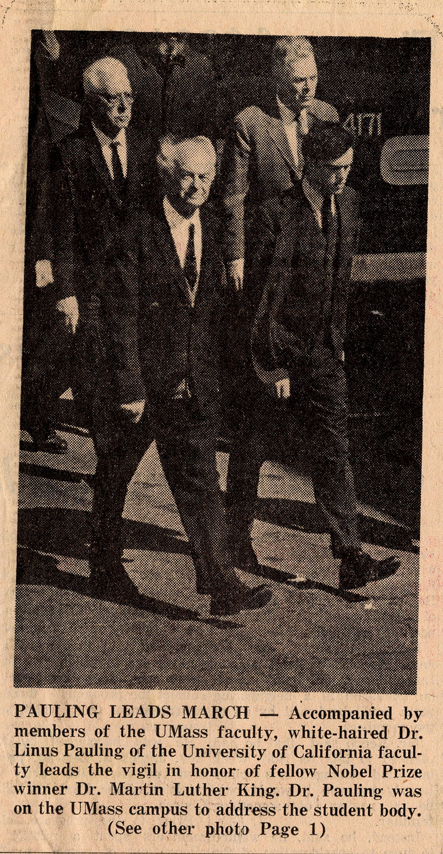
Pauling happened to be at the University of Massachusetts a mere five days after Martin Luther King Jr. was assassinated. Invited to deliver a series of lectures as the university’s first Distinguished Professor, Pauling fashioned his remarks around the topic of the human aspect of scientific discoveries. Reflecting on the tumult of the previous week, Pauling told his audience that it was not enough to mourn the fallen civil rights leader. Rather, individuals of good conscience were obligated to carry King’s legacy forward by continuing the work that he began.

In keeping with this theme over the course of his lectures, Pauling emphasized the scientist’s responsibility to ensure that discoveries be used for the good of all humanity and society, rather than in support of war and human suffering. Scientific inquiry should also emphasize solutions to current issues, he felt, pointing to the lack of equality in access to medical care in the United States as one such issue. Pauling saw his work in orthomolecular medicine as potentially solving this problem: vitamins were fairly inexpensive, more accessible, and could, he believed, significantly improve one’s mental and physical well-being.


1968s.5

Notes used by Pauling for his talk, “The Scientific Revolution,” delivered as a component of the lecture series, “The Revolutionary Age, the Challenge to Man,” March 3, 1968.

Pauling made similar connections to his work on sickle cell anemia.

Though he was no longer involved in the daily operations of the CSDI, he continued to participate in a public lecture series that the center sponsored throughout his time in San Diego. In one contribution to a series titled “The Revolutionary Age: The Challenge to Man,” Pauling put forth a potential solution to sickle cell disease. As science had succeeded in identifying the gene mutation responsible for the disease, Pauling believed that forms of social control could be used to prevent carriers of the mutation from marrying and procreating. Over time, Pauling reasoned, the mutation would eventually be phased out.

Pauling specifically called for the drafting of laws that would require genetic testing before marriage. Should tests of this sort reveal that two heterozygotes (individuals carrying one normal chromosome and one mutation) intended to marry, their application for a license would be denied. Pauling put forth similar ideas about restricting the number of children that a couple could have if one parent was shown to be a carrier for sickle cell trait.

In proposing these ideas, Pauling aimed to ensure that his discovery of the molecular basis of sickle cell disease was used to decrease human suffering. Likewise, he felt that whatever hardships the laws that he proposed might cause in the short run, the future benefits accrued from the gradual elimination of the disease would justify the legislation.

Partly because he called this approach “negative eugenics,” Pauling came into harsh criticism for his point of view; indeed, his ideas on this topic remain controversial today. In a number of the lectures that he delivered around the time of his CSDI talk, however, Pauling took pains to clarify that his perspective was not aligned with the broader field of eugenics, a body of thought to which he was opposed. On the contrary, Pauling’s focus was purely genetic and his specific motivation was borne out of a desire to eliminate harmful genetic conditions.


Bruno

Bruno Zimm. Credit: University of California, San Diego

At the end of February 1968, Pauling turned 67 year old, and the University of California regents used his age as a mechanism to hold up discussions about his obtaining a permanent appointment in San Diego. Sixty-seven, the board argued, was the typical retiring age within the UC system. Moreover, the UC regents were empowered to veto any age-related retirement exceptions and, given his radical political views, Pauling was unlikely to receive any support at all from the group, much less an exception.

One of the stated reasons why the regents harbored concerns about Pauling’s politics was his increasingly strident rhetoric. Pauling frequently commended student strikes and demonstrations, and although he emphasized nonviolence as the most effective means to foster social change, he encouraged students to recognize that authorities may incite violence through tactics of their own. In these cases, he felt that retaliation was justified, even necessary.

Pauling also believed that the regents and their trustees wielded too much power; for him they were part of a system that largely inhibited social progress and took power away from students. For their part, the regents saw Pauling in a similar light: a dangerously powerful radical who was constraining the university’s capacity to grow.

Realizing that, in all likelihood, Pauling was soon to be forced out, his UCSD colleagues Fred Wall and Bruno Zimm began searching for a way to shift the governing authority for his reappointment to the university president, Charles Hitch, with whom Pauling had maintained a positive relationship. After months of negotiations, Zimm succeeded in winning for Pauling a second year-long appointment.

Pauling expressed gratitude to Zimm for his efforts, but the slim possibility of a permanent position at UCSD had emerged as a source of lingering dismay. Looking for a longer term academic home, Pauling began considering other universities that might also provide better support for his research.

Over time, Ava Helen had also found herself frustrated with UCSD and La Jolla in general. In particular, she disliked their rental house and missed their previous home in Santa Barbara, where she had been able to tend a beautiful garden. As 1968 moved forward, the couple began spending more and more time at Deer Flat Ranch, with Ava Helen hinting that she would like to make the ranch their permanent home in the coming years.

Dr. Pauling’s Prediction of a Mutation in Beta-Globin Which Causes Sickle Cell Anemia and How This Prediction Impacted My Research

[Guest post written by John Leavitt, Ph.D., Nerac, Inc., Tolland, CT.]

Linus Pauling lecturing on sickle cell anemia, Kyoto, Japan. 1955

In September 2010, the company BlueBird Bio announced that it had cured a patient with the hemoglobinopathy, beta-thalessemia, by correcting the genetic defect in beta-globin that this patient inherited from his parents. This came 61 years after Linus Pauling and his associate, Harvey Itano, explained the cause of hemoglobinopathies such as sickle cell anemia. Beta-thalessemia like sickle cell anemia is caused by an inherited mutation in the beta-globin gene, just a different mutation. In the case of thalessemia, the defective beta-globin gene product disappears, whereas the defective beta-globin in sickle cell anemia remains stable to wreak havoc on the body. BlueBird Bio accomplished this first cure of a hemoglobinopathy by removing the blood-forming hematopoietic stem cells from the patient, engineering his cells ex vivo with a correct beta-globin gene, and then putting the cells back into the patient. The stem cell transplant sustained itself and produced red blood cells which functioned normally in the circulatory system. For the first time in this patient’s 18 year-old life, he did not have to have a monthly blood transfusion.


In late September 1981, when I gave a seminar at the Pauling Institute of Science and Medicine in Palo Alto, CA, I noticed that Dr. Pauling was smiling during the talk. He was aware of the discovery of the muscle isoform of actin by his friend Albert Szent-Györgyi and knew about the structure and function of actins (the subject of my talk). After reading Dr. Pauling’s 1949 paper on the molecular nature of the sickle cell trait, I understood that he was seeing during my talk the very same experiments in my discovery of a mutant human beta-actin that he and Harvey Itano had performed, which led to the prediction of a mutation in the hemoglobin protein that caused sickle cell anemia. His paper was the very first to describe the molecular genetic basis of a human disease. By 1981 there was plenty of conceptual evidence to suggest how I could look for mutations in proteins using electrophoretic separation of complex mixtures of cellular proteins. In 1949 though, Dr. Pauling was way ahead of his time. In his and Itano’s case, the plan was well thought out based upon years of characterization of oxygen bonding to the heme of the globin molecule. By contrast, I was very lucky to find a mutation in the most abundant structural protein of the cell, cytoskeletal actin in a human fibrosarcoma cell.

Harvey Itano.

It was probably evident in 1949 that hemoglobin amounts to about 95 percent of the total protein of a mature red blood cell; so these cells were essentially bags of hemoglobin molecules – globin polypeptides with attached heme moieties with an iron atom that bound oxygen. The heme-bound iron carries oxygen through the arterial system to cells for respiration. After delivery of oxygen to tissues, these red blood cells (RBCs) return carbon dioxide to the lungs through the venous system for expiration. In sickle cell anemia, after RBCs deliver oxygen throughout the body, the RBCs take on a sickled shape, clog the venous system and lyse, causing a wide variety of systemic problems. Pauling and Itano predicted that this change in RBC architecture was a direct consequence of “two to four” charged amino acid changes in the globin complex, which consists of two beta-globin subunits and two alpha-globin subunits (this was not known then). Because of the science that came after their discovery, we know now that the genetic mutation in the beta-globin moiety is a single amino acid exchange of glutamic acid to valine resulting from a single nucleotide transition (A to T transition) in codon 6 of the beta-globin gene encoding the 147 amino acid polypeptide. Thus two positive charges were added to the hemoglobin molecule by this mutation. Pauling and Itano concluded that these charge alterations caused RBC sickling.

Important discoveries can be quite simple. The figure below is the key experiment carried out by Pauling and Itano, an electrophoretic separation of hemoglobin based upon its isoelectric point (net charge). Because of the mutation in codon 6 present in both inherited beta-globin alleles, the hemoglobin complex migrated to the right of the normal hemoglobin by approximately “two to four” positive charges (panel B compared with panel A). At pH 6.9 the normal hemoglobin was shown to have an isoelectric point of 6.87, migrating as a negative ion, whereas the mutated hemoglobin had an isoelectric point of 7.09 migrating as a positive ion. We know now that this electrophoretic change in the hemoglobin complex described by Pauling and Itano is due to the loss of a single negative charge in a glutamic acid residue (replaced with an uncharged valine residue) near the N-terminus of the two beta-globin moieties of the hemoglobin molecule. Today, the fact remains that this is the only mutation in hemoglobin that causes sickle cell anemia, although other beta-globin mutations cause other hemoglobinopathies like beta-thalessemia. Panel C shows the electrophoretic behavior of hemoglobins in a heterozygous carrier of the disease-causing mutation (Panel D is a control mixture of the globins in panels A and B). Much more insight about these phenomena is discussed in the Pauling and Itano paper but the charge alteration in hemoglobins is the basic observation.

Pauling experiment

(click to enlarge)

Fast-forward to 1976. I decided to look for evidence of charge-altering mutations in a protein profile of about 1,000 visible proteins (polypeptides) comparing normal and neoplastic cells by looking for Pauling and Itano’s evidence of mutations, e.g. minor charge alterations in proteins in the protein profile. A technique had just been developed by Patrick O’Farrell which permitted high-resolution separation of virtually all major protein gene products of the cell.

An elegant study was performed by Greg Milman at the University of California at Berkeley who demonstrated that one could predict the occurrence of mutations in the relatively minor protein, the enzyme hypoxanthine phosphoribosyltransferase (HPRT), in HeLa cells by the positional changes in the HPRT polypeptide in high-resolution two-dimensional polyacrylamide gels within complex profile of proteins separated both by their charge (isoelectric point) and their molecular weight. When I saw Milman’s result I decided to use this technique to compare normal and neoplastic human cells to see if I could identify charge alterations similar to those demonstrated by Pauling and Itano in hemoglobin and by Milman in HPRT.

I labeled the proteins of normal and tumor-forming human fibroblasts with S35-methionine and separated them using O’Farrell’s two-dimensional technique (isoelectric point separation is a tube gel followed by molecular weight sieving in an SDS slab gel). Then I fixed the proteins in the two-dimensional slab gel and stained these proteins with Coomassie blue dye.

mutant actin further annotated

With the dye you could only see the most abundant proteins and I was surprised to see this pattern of actins in the tumor-forming fibroblasts shown above. This image is actually the image of the radioactive protein pattern in the region of actins (pI 5.3 to pI 5.1, molecular weight Mr about 42,000) developed after a very short autoradiographic exposure to X-ray film (a digital image). Normally you only see one beta-actin spot barely separated from the gamma-actin spot. Gamma actin is a second isoform of actin encoded by a separate gene which differs by only four amino acids from beta-actin. Normally there is about twice as much beta-actin at isoelectric point 5.2 as gamma actin and both actins together amount to 5-10 percent of the total cellular protein. But half of the normal beta-actin was missing and a new more negative spot at isoelectric point 5.3 appeared. I was able to show that this was a new form of beta-actin by tryptic peptide separation and other criteria. The observation that the new variant migrated slower in the second dimension as a larger protein was later attributed to a frictional effect in the gel sieve due to an altered conformation caused by the amino acid change.

These observations and other differences in protein expression between the normal and tumor-forming fibroblasts were published in the Journal of Biological Chemistry in February 1980. A second paper was published a month later demonstrating that a T-cell leukemia also had a beta-actin anomaly which suggested loss of a beta-actin allele. It is now well established that reorganization of the actin cytoskeleton occurs when cells become cancerous, although mutations in the structural gene may be less common. These alterations can also be caused by changes in actin-binding proteins.

Later in the year, with my colleagues at the Max-Planck in Goettingen Germany, Klaus Weber and Joel Vandekerckhove, I published the sequences of the normal human beta- and gamma-actins and the mutant beta-actin in Cell. The normal and mutated sequence of human beta-actin is shown in the figure below.

The simple electrophoretic difference between the mutant and normal beta-actin was a single amino acid exchange of a neutral glycine for a negatively charged aspartic acid at amino acid residue 244 in the 374 amino acid polypeptide chain, an observation similar to Pauling and Itano’s hypothesis 32 years earlier. An amino acid exchange at this residue in the actin polypeptide chain had never been observed in any eukaryote. Two years later I cloned the mutant and wildtype human beta-actin genes at the Pauling Institute and formally proved the existence of the mutation at the level of the gene. This mutation was caused by a single nucleotide change in the gene. Several years later my colleagues and I demonstrated that acquisition of this simple mutation contributed to the tumorigenic phenotype of the cells in which it arose.

actin sequence with arrow

The sequence of human beta-actin and its amino acid 244 mutation (the most highly conserved protein in eukaryotes).


Ed Note: This week marks the sixth anniversary of the creation of the Pauling Blog.  Birthed to help promote the unveiling of a postage stamp, the blog, 461 posts later, has developed into a resource of consequence with an audience that is steadily growing.  For those who might be interested in how the project operates, please see this post that we ran one year ago.

As always, we thank you for your continued readership.  We plan to keep researching and writing, so please keep coming back!

1955i.45

An Era of Discovery in Protein Structure

Linus and Ava Helen Pauling, Oxford, 1948.

[The Paulings in England: Part 4 of 5]

Though metals were consuming a good portion of his time during his fellowship at Oxford, Linus Pauling’s other projects never strayed far from his thoughts.  High on the list were the mysteries of proteins, whose structures and functions were slowly starting to be unraveled.

Pauling’s interest in proteins was spurred in the mid-1930s when the Rockefeller Foundation began to look most favorably upon the chemistry of life when deciding where their grant money would go. Early on, Pauling set out to tackle hemoglobin and though his affair with the molecule lasted for the remainder of life, Pauling certainly didn’t limit himself to the study of just one protein.

At a time when most were looking at proteins from the top down, trying to sort out the complicated data produced by an x-ray diffraction photograph of an entire protein, Pauling was working from the bottom up, in the process determining the structures of individual amino acids – the building blocks of proteins.

A specific protein that kept coming back into view over the years was keratin. In the 1930s, the English scientist William Astbury had studied the structure of wool, which along with hair, horn, and fingernail is made up primarily of this enigmatic protein, keratin. Astbury proposed that the structure was akin to a flat, kinked ribbon, but Pauling disagreed. “I knew that what Astbury had said wasn’t right,” Pauling recalled, “because our studies of simple molecules had given us enough knowledge about bond lengths and bond angles and hydrogen-bond formation to show that what he said wasn’t right. But I didn’t know what was right.” Pauling attempted to construct a model at the time, but could not match his structure to the measurements dictated by Astbury’s blurry x-ray diffraction images. Pauling wrote the project off as a failure and continued pursuing other interests.

In 1945 Pauling found himself seated next to Harvard medical Professor William B. Castle on a railroad journey from Denver to Chicago. Castle was a physician working on the nature of sickle cell anemia and the conversation that he shared with Pauling planted a seed in Pauling’s mind about the cause of this debilitating disease.

In the bodies of those suffering from sickle cell anemia, red blood cells assume a sickled shape when they are in the deoxygenated venous system but retain their normal flattened disk shape in the oxygen-rich arterial system. Noting this, Pauling suggested that perhaps the source of the problem could be a defect in the oxygen-carrying protein itself: hemoglobin.

Amidst his travels in Europe, Pauling continued to act on this idea as maestro from afar, directing the scientists in his Caltech laboratory to continue searching for differences in the hemoglobin of normal and sickled cells. In the meantime, he sought out and communicated new ideas gleaned from meetings such as the Barcroft Memorial Conference on Hemoglobin, held at Cambridge in June 1948. Pauling’s research team, in particular Harvey Itano and S. Jonathan Singer, were able to show experimentally that his hunch had been right, and less than a year after his return to Pasadena a paper was published that established sickle cell anemia as the first illness to be revealed as a truly molecular disease.

Linus and Peter Pauling at the model Bourton-on-the-water, England. 1948.

While in England, Pauling had occasion to interact closely with a number of scientific greats.  Among these were his close friend Dorothy Crowfoot Hodgkin, who is credited as a pioneer in the development of protein crystallography and was the winner of the 1964 Nobel Prize for Chemistry.  Likewise, Pauling conversed with Max Perutz, a protege of Sir William Lawrence Bragg‘s at the Cavendish Laboratory at Cambridge, who would go on to discover the structure of hemoglobin and receive the Nobel Prize for Chemistry in 1962.  While fruitful in many respects, these interactions served to increase Pauling’s feelings of urgency as concerned the race to determine the structure of proteins.

Bragg shared the 1915 Nobel Prize in Physics with his father for their early development of X-ray crystallography, and though there existed a long-standing scientific rivalry between Pauling’s and Bragg’s laboratories, it wasn’t until Pauling saw, with his own eyes, the work that was being done that he admitted he was “beginning to feel a bit uncomfortable about the English competition.” As he wrote to his colleague Edward Hughes back at Caltech

It has been a good experience for me to look over the x-ray laboratory at Cambridge. They have about five times as great an outfit as ours, that is, with facilities for taking nearly 30 x-ray pictures at the same time. I think that we should expand our x-ray lab without delay.

This realization prompted Pauling to get researchers in his lab started on work with insulin – an arduous and complicated process that required sample purification and crystallization prior to x-ray investigation. In relaying research findings from English scientists working on insulin to his partners back in Pasadena, Pauling intimated that

It is clear that there is already considerable progress made on the job of a complete structure determination of insulin. However, there is still a very great deal of work that remains to be done, and I do not think that it is assured that the British school will finish the job. I believe that this is the problem that we should begin to work on, with as much vigor as possible, under our insulin project.

Little did Pauling know that, while laying in bed, using little more than a piece of paper, a pen and a slide rule, he would soon make a major breakthrough in protein chemistry on his own.

The Medical Research of Linus Pauling

By Tom Hager

[Ed Note:  In October 2010, Pauling biographer Tom Hager delivered a talk sponsored by the Oregon Health Sciences University which detailed and discussed the various contributions that Linus Pauling made to the medical sciences, including the controversy over his strong interest in orthomolecular medicine.   With the author’s permission, excerpts of this talk are being presented on the Pauling Blog over the next three posts.  The full text of Hager’s OHSU lecture is available here.  Those with an interest in learning more about Hager’s work, including his latest research on food issues and world hunger, are encouraged to visit his blog at http://thomashager.net.]

[Part 1 of 3]

Oil portrait of Linus Pauling, featuring a model of the alpha-helix in the foreground. 1951. Portrait by Leon Tadrick.

By 1939, at the age of 38, Linus Pauling was a full professor and head of the chemistry division at Caltech, as well as the father of four children (three sons, Linus, Jr., Peter, and Crellin; and a daughter, Linda).

He was also beginning to turn his considerable talents toward understanding the complicated molecules inside the human body. He started with proteins.

The Molecules of Life

Determining the structure of proteins at this time was a gigantic problem. Most were difficult to purify, easily degraded, and hard to characterize. Proteins appeared to be not only gigantic, comprising hundreds or thousands of atoms – much too large to solve directly with x-ray crystallography – but also relatively fragile, losing their function (denaturing) after even slight heating or mechanical disturbance. No one at the time was even sure that they were distinct molecules – one popular theory held that proteins formed amorphous colloids, gels that did not lend themselves to molecular study.

Studying them at the molecular level seemed an impossible task with the tools available in the late 1930s. But Pauling took on the challenge. He started with the building blocks of proteins, the amino acids, and directed his growing lab team toward pinning down their precise structures. Then he set himself to figuring out how they formed protein molecules, often building models out of wood, wire, and paper.

He based his approach in part on the ideas of the German biochemist Emil Fischer. Like Fischer, Pauling came to believe that proteins were long molecular chains of amino acids linked end-to-end. Working with Alfred Mirsky in the mid-1930s, Pauling discovered that the denaturing of proteins resulted from breaking weak bonds, called hydrogen bonds, that pinned these chains into specific shapes. Between the early 1930s and early 1950s he made a string of important discoveries about hemoglobin, antibodies (including the most sophisticated work at the time into the structural relationship between antibody and antigen), enzymes, and other proteins.

Foldable paper model of the alpha-helix protein structure published in the Japanese journal Chemical Field, 1954.

In May 1951, he put everything he knew into a celebrated series of seven papers detailing the structures of a number of proteins at the level of individual atoms, including the structure of the single most important basic form of protein, the alpha helix (a hydrogen-bonded helical chain that is a structural component of almost every protein). It was an astounding breakthrough, and it opened the door for an understanding of biology at the molecular level. Within two years, Watson and Crick had used his approach to decipher the structure of DNA.

Biological Specificity

But structure was not everything. Pauling realized that life resulted not from individual molecules, but from the interactions between them. How did organisms make offspring that carried their specific characteristics? How did enzymes recognize and bind precisely to specific substrate molecules? How did antibodies recognize and bind to specific antigens? How did proteins, these flexible, delicate, complex molecules, have the exquisite ability to recognize and interact with target molecules?

It all fell under the heading of biological specificity at the molecular level. Pauling directed much of his attention here during through the 1940s, performing a great deal of careful work on the binding of antigens to antibodies.

Drawings of antibodies and antigens made by Linus Pauling in the 1940s.

His findings were surprising. Pauling demonstrated that the precise binding of antigen to antibody was accomplished not by typical chemical means – that is, through covalent or ionic bonds — but solely through shape. Antibodies recognized and bound to antigens because one fit the other, as a glove fits a hand. Their shapes were complementary. When the fit was tight, the surfaces of antibody and antigen came into very close contact, making possible the formation of many weak links that operated at close quarters and were considered relatively unimportant in traditional chemistry — van der Waals’ forces, hydrogen bonds, and so forth. To work, the fit had to be incredibly precise. Even a single atom out of place could significantly affect the binding.

Having demonstrated the importance of complementary structure with antibodies, Pauling extended his idea to other biological systems, including the interaction of enzymes with substrates, odors with olfactory receptors, and to the possibility of complementary structure in genes.

Pauling’s idea that biological specificity was due in great part to complementary “fitting” of large molecules to one another proved to be essential in the development of molecular biology. His research now formed a coherent arc, from his early work on the chemical bond as a determinant of molecular structure, through the structures of large molecules (first inorganic substances, then biomolecules), to the interactions between large biomolecules.

He carried out much of this research during World War II, when he also worked on synthetic plasma substitutes and a fruitless search for ways to produce artificial antibodies.

He had already earned a place among the nation’s leading researchers in the medical applications of chemistry. But his greatest triumph was still to come.

Sickle-Cell Anemia

Toward the end of World War II, Pauling’s reputation was great enough to earn him an invitation to join a national committee that was brainstorming the best structures for postwar medical research. This committee’s work led to the foundation of the National Institutes of Health.

Pauling was the only non-physician asked to join the committee.

At a dinner with other members one night, talk turned to a rare blood disorder called sickle-cell anemia. One of his dinner companions described how red blood cells in the victims were twisted into sickle shapes instead of discs. The distortion appeared to hinder the blood cells’ transport through capillaries, resulting in joint pain, blood clots, and death. The disease primarily affected Africans and African Americans. What caught Pauling’s attention most, however, was one odd fact: Sickled cells appeared most often in venous blood, rather than in the more oxygenated blood found in the arteries.

Pastel drawing of sickled Hemoglobin cells, 1964. Drawing by Roger Hayward.

He thought about this during the next few days. From his previous work with blood, he knew that red cells were little more than bags stuffed with hemoglobin. He had also shown that hemoglobin changed its shape slightly when it was oxygenated. If the red cells were changing shape, perhaps it was because the hemoglobin was altered in some way. What if the hemoglobin molecules in sickle-cell patients were changed in some way that made them clump, stick to one another, as antigens stick to antibodies? Perhaps something had changed that made the hemoglobin molecules complementary in shape. Perhaps adding oxygen reduced the stickiness by changing the molecules’ shape.

He presented his ideas as a research problem to Harvey Itano, a young physician who was then working on his Ph.D. in Pauling’s laboratory. Itano, later joined by postdoctoral fellow John Singer, worked for a year trying to see if sickle-cell hemoglobin was shaped differently from normal hemoglobin. They found no detectable differences in any of the tests they devised. But they kept at it. Finally, in 1949, using an exquisitely sensitive new technique called electrophoresis that separated molecules by their electric charge, they found their answer: Sickle-cell hemoglobin carried more positive charges on its surface.

This was an astounding discovery. A slight change in the electrical charge of a single type of molecule in the body could spell the difference between life and death. Never before had the cause of a disease been traced to a molecule. This discovery – to which Pauling attached the memorable title “molecular disease” – received widespread attention. Itano and Singer’s followup work demonstrated the pattern of inheritance for the disease, firmly wedding molecular medicine to genetics.

Medical Chemistry

It was a great triumph – there was talk of a Nobel Prize in Medicine or Physiology for Pauling – and it led Pauling to make greater efforts in the medical field. He encouraged M.D./Ph.D. candidates, hired physicians to work in his laboratory, and began focusing his own research on medical problems, including developing a new theory of anesthesia.

He was ahead of his time. An example of what the atmosphere was like: Pauling noted that as he went around in the late 1940s seeking funds for a comprehensive marriage of biology and chemistry to attack medical problems, people at funding agencies were telling him that they found the term “medical chemistry” to be “a disturbing description.”

“Mental Deficiency & Brain Chemistry.” May 1, 1964.

In the late 1950s, Pauling extended his concept of molecular disease to the brain. After reading about phenylketonuria (PKU) – a condition in which a mental defect can be caused by the body’s inability to metabolize an amino acid, phenylalanine, leading to a buildup of that substance and others in the blood and urine – Pauling theorized that the problem might be caused by a defect in an enzyme needed to break down phenylalanine. PKU, in other words, might be another molecular disease. Now interested in the possibility that there might exist a range of molecular mental defects, Pauling visited a local mental hospital, saw other patients whose diseases seemed hereditary, and decided to seek support for an investigation into the molecular basis of mental disease. The Ford Foundation in 1956 awarded him $450,000 for five years’ work – a vindication of Pauling’s approach and a tribute to his reputation. The grant, however, yielded little in the way of immediate results, with much of the funding going toward testing his (ultimately found to be mistaken) theory of anesthesia.

The long-term results were more significant. Pauling’s immersion in the field, thanks to the Ford grant, led him to read widely in psychiatry and general health, always on the lookout for another molecular disease that might lend itself to new therapy. By the mid-1960s he was coalescing his findings into another overarching theory, this one combining much of what he knew about chemistry and health. He called his new idea “orthomolecular” medicine.

Remembering Harvey Itano

Portrait of Harvey Itano, 1954. Image courtesy of the Caltech Institute Archives.

“The discovery by Dr. Itano of the abnormal human hemoglobins has thrown much light on the problem of the nature of the hereditary hemolytic anemias, and has changed these diseases from the status of poorly understood and poorly characterized diseases into that of well understood and well characterized diseases.”

-Linus Pauling, 1955.

We were saddened to learn of the death of Harvey A. Itano, emeritus professor of pathology at the University of California, San Diego.  Dr. Itano passed away on May 8, 2010 at the age of 89.

Best known professionally for his work on sickle cell anemia, Itano’s early personal history makes for fascinating reading.  According to this excellent obituary issued by UCSD

Itano was born in Sacramento, CA on November 3, 1920, the oldest of four children of Masao and Sumako Itano, originally of Okayama-ken, Japan.  A star student at UC Berkeley, he graduated in 1942 with highest honors in chemistry.  He was unable to attend his own graduation ceremony, because he and his family were confined to internment camps established after the bombing of Pearl Harbor for the detention of Japanese and Japanese-Americans living in the western US.  In recognition of his outstanding achievements as a student, having earned the highest academic record in his class, then-UC President Robert Gordon Sproul personally awarded him the University Medal during his internment.

[…] He was released from the camp on July 4, 1942, the first of the Nisei (second generation Japanese-Americans) to be released to attend colleges and universities.  He attended the St. Louis School of Medicine, where he earned his MD in 1945 before continuing his studies at California Institute of Technology, earning a PhD in Chemistry and Physics in 1950.

It was at Caltech that Itano came into contact with Linus Pauling, his major professor during his doctoral studies and research colleague for the duration of a four year post-doctoral stint in Pasadena.  Over the course of this time period, Itano, Pauling and their collaborators made a series of significant contributions to the field of molecular biology.

Most prominent among these contributions was a 1949 paper published in Science, titled “Sickle Cell Anemia, A Molecular Disease.”  Authored by Pauling, Itano, S. Jonathan Singer and Ibert C. Wells, the paper presented experimental evidence in support of Pauling’s theory that sickle cell anemia could be traced to significant abnormalities in the hemoglobin molecules of those suffering from the disease.  The paper was quickly recognized to be the first solid proof of the existence of a “molecular disease.”

In his book Force of Nature, Pauling biographer Thomas Hager comments on the importance of this discovery.

People had theorized in broad terms about the molecular basis of disease before, but no one had ever demonstrated it the way Pauling’s group did….By pinpointing the source of a disease in the alteration of a specific molecule and firmly linking it to genetics, Pauling’s group created a landmark in the history of both medicine and molecular biology.

Itano spent much of his long career furthering the breakthroughs signaled in the 1949 paper.  Among other achievements, he developed a “rapid diagnostic test” for sickle cell anemia which would quickly indicate whether or not a given blood sample would sickle.  With S. J. Singer, Itano also described the condition of sicklemia, an intermediate and less severe stage of sickle cell anemia in which a patient’s blood contains a mix of normal hemoglobin and sickled hemoglobin cells.

Harvey Itano and Linus Pauling. 1980s.

Linus Pauling held Itano in high regard, both as a scientist and as a person.  In a lengthy award nomination that Pauling composed for Itano in 1955, Pauling describes the specifics of Itano’s contribution to the team’s molecular disease breakthrough while noting his “great natural ability and thoroughly sound training in chemistry and related sciences as well as in medicine.”  Of the man, Pauling wrote

His success must also be attributed in part to his excellent personality.  He is quiet and pleasant in manner, and is well liked by all of his associates.  During his eight years at the California Institute of Technology he made many friends, and he was uniformly successful in effective collaboration with a number of co-workers.  He is original, clearheaded, keen, and critical in his scientific work.

Itano maintained a keen interest in his rich genealogical background, and those who wish to learn more about his story are encouraged to visit the Itano family history website.  A great deal more about Itano’s role in the sickle cell anemia and molecular disease story is likewise available at It’s in the Blood!  A Documentary History of Linus Pauling, Hemoglobin and Sickle Cell Anemia.

Mastering Genetics: Pauling and Eugenics

Illustration from Medical World News article,

Illustration from Medical World News article, “Sickle Cell Anemia” December 3, 1971.

“I have suggested that the time might come in the future when information about heterozygosity in such serious genes as the sickle cell anemia gene would be tattooed on the forehead of the carriers, so that young men and women would at once be warned not to fall in love with each other.”
-Linus Pauling, August 15, 1966

After declaring sickle-cell anemia to be a “molecular disease” in the late 1940s, Pauling spent more than a decade describing the cause of the disease and the significance of its unique origins to his fellow academics. Unfortunately, though interested, his colleagues seemed more concerned with the concept of a molecular disease than its real world application in genetics and medicine. Beginning in 1958, Pauling became a vocal advocate of genetic counseling, focusing especially on sickle-cell anemia among the African American population. His efforts went largely unnoticed by both researchers and the general public alike.

Frustrated with his unsuccessful endorsement of genetic counseling, Pauling chose to take his ideas a step further. In 1962, Pauling began a public campaign in support of negative eugenics – the restriction of human breeding and childbirth as a means of minimizing the sharing of hereditary diseases. He advocated genetic testing as a requirement for obtaining a marriage license. Perhaps even more controversial, Pauling recommended placing legal restrictions on marriage and childbirth between carriers of hereditary diseases.

Listen: Pauling on Marriage Tests and the Disclosure of Genotype Information

Pauling recognized the difficulty of controlling and monitoring a program of this magnitude. Without being able to easily identify carriers of various diseases, the public could not effectively choose sexual partners, thus lessening the potential effectiveness of employed eugenics. As a solution, in the late 1960s, Pauling began suggesting a means of visibly marking disease carriers – a tattoo on the forehead, clearly marking the individual as the carrier of a specific disease. Not surprisingly, this suggestion engendered a great deal of criticism. He was compared with the likes of Hitler by his critics who drew parallels between the proposed tattoo and the yellow star worn by Eastern European Jews during the reign of the Nazi party.

In reflecting upon Pauling’s stance, it is important to note that he was not interested in positive eugenics – the manipulation of genetic combinations as a means of developing a superior human. Rather, he intended only to minimize human suffering and found the idea of building a “super race” highly undesirable. Pauling was also a critic of the concept of genetic purity. He was concerned with purifying the human gene pool of harmful diseases, but he was not motivated by the desire to manipulate intelligence, appearance, strength, etc.

Pauling insisted that his ideas, though extreme, were meant to decrease human suffering rather than to segregate and belittle. Though Pauling faced many critics, he did have supporters as well. Nobel laureate Sir Peter Medawar agreed with Pauling, famously stating,

It is humbug to say that such a policy violates an elementary right of human beings. No one has conferred upon human beings the right knowingly to bring maimed or biochemically crippled children in the world.

During the 1960s, Pauling’s critics began discussing the effect that negative eugenics could have on evolution. Roderic Gorney, a psychiatrist, argued that over a long enough period of time, eugenics could redirect and even supersede the process of natural selection.

For example, consider the effect of negative eugenics in relation to sickle-cell anemia. An individual with sickle-cell anemia has two sickle-cell alleles. Typically, sufferers of sickle-cell anemia are plagued by a host of related health problems, often leading to an early death. Some individuals, however, possess only one sickle-cell allele. These individuals exhibit some sickling of the blood cells, but are otherwise able to live normal, healthy lives. Because sickle-cell anemia is a hereditary disease, it is passed on in Mendelian fashion. As a result, a person with a single sickle-cell allele, when paired with a healthy individual, has a 25% chance of giving birth to a child with one sickle-cell allele. When paired with another single-trait individual, there exists a 50% chance that a child will have one sickle-cell trait, and a 25% chance that the child will be afflicted with full sickle-cell anemia.

Gorney argued that sickle-cell anemia, if left alone, would eventually be removed from the human gene pool. He explained that, because individuals suffering from sickle-cell anemia rarely live to procreate, few instances of sickle-cell anemia are added to the collective gene pool. Similarly, a single-allele individual has a statistical opportunity to produce children with sickle-cell anemia when paired with another carrier. These offspring will die at a young age, further reducing the number of carriers present in the next generation. As a result, over a period of time, the number of sickle-cell carriers would decrease to nothing.

Negative eugenics, however, allows sickle-cell carriers to identify other carriers and instead mate with healthy individuals, producing more children with a single sickle-cell allele. If this process were to continue indefinitely, more and more humans would be heterozygous for sickle-cell anemia, rendering it virtually impossible for natural selection to remove the disease from the human gene pool. This argument could, in fact, be applied to any similar hereditary disease.

“Bad Genes and Marriage,” New York Post, October 21, 1968.

Pauling acknowledged Gorney’s concerns but countered that, without eugenics, preventative medicine would have a much more damaging effect. Pauling felt that modern medicine (antibiotics, chemotherapy, prescription drugs, etc.) helped prolong the lifespan of sick or diseased individuals, sometimes allowing them to procreate and pass along hereditary diseases. As such, modern medicine was effectively undoing natural selection, leaving negative eugenics as the best hope for maintaining a balanced, healthy population.

In the early 1970s, Pauling began to run into trouble. His main focus throughout his eugenics campaign was the elimination of sickle-cell anemia, a disease that had originated in Africa where it became common among the native population because of its ability to prevent malaria. When slave traders brought African captives to North America, sickle-cell anemia was introduced to the United States. Due to racial segregation and the social mores that developed in the U.S. over the intervening 300 years, very few individuals outside of the African American population were afflicted with sickle-cell disease. For these reasons, Pauling advocated blood testing among the African American population. As the Civil Rights movement gained momentum, Pauling’s suggestions were seen as racist, and even as an attempt to cast African Americans as genetically inferior and meriting legal restrictions on their rights to marriage and procreation.

Frustrated and embarrassed by the criticism that he was receiving, Pauling fell silent on the topic of eugenics. In the past, when faced with heavy opposition, Pauling had always held his ground. But this episode was different. By the end of 1972, Pauling had given up his negative eugenics campaign and turned to other means of improving the human condition.

For more information on Pauling and his work with genetics, visit “It’s in the Blood! A Documentary History of Linus Pauling, Hemoglobin and Sickle Cell Anemia” or Linus Pauling Online.