Normal Expression of Human Beta-Actin (Cloned at LPISM) Acts as a Tumor Suppressor – A Novel Hypothesis

[Guest post written by John Leavitt, Ph.D., retired Senior Scientist at LPISM in Palo Alto CA from 1981 to 1988; living in Woodstock CT.]

leavitt2016

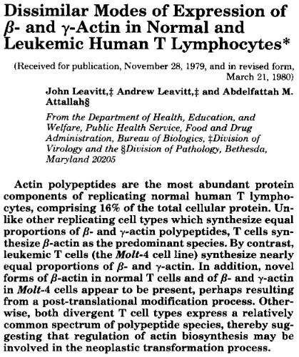
In 1980, Klaus Weber at the Max-Planck Institute and I published the amino acid sequence of human beta- and gamma-cytoplasmic actins. In 1981, after we completed this work, Klaus asked me “What are you going to do next?” I told him that I was moving to the Linus Pauling Institute of Science and Medicine in Palo Alto, California, and that I was going to clone the human beta-actin gene. My reason was that I had discovered a mutation in beta-actin that was associated with a tumorigenic human fibrosarcoma cell line. I wanted to test the hypothesis that this mutation contributed to the tumorigenic potential of this fibrosarcoma.

In 1984, I published the cloning of multiple copies of both the normal (wildtype) human beta-actin gene and multiple copies of the mutant gene. These actins are the most abundant proteins of all replicating mammalian cells and most other cells, down to yeast. (My story of meeting Dr. Pauling, moving from the National Institutes of Health to the LPISM, and our work on the role of this actin mutation in tumorigenesis in our model system was recounted in an article posted at the Pauling Blog in 2014.) In 2013, Schoenenberger et al. at the Biozentrum in Basel, Switzerland, reproduced all of our findings in a different cell system, a rat fibroblast model system, and extended our findings (see our review of their work).

A year ago, in June 2015, Dugina et al published a paper that proposed that altering the ratio of these two actins regulated either suppression or promotion of cancerous cell growth (more work needs to be done). I was very surprised by this idea – even though our work at LPISM had suggested this, I hadn’t thought of putting our observations into the language of “tumor suppression” and “tumor promotion.” Perhaps this was because, in the 1980s, hundreds of so-called “oncogenes” (tumor promoters) and tumor suppressor genes were being cataloged, and our findings were suggesting that a so-called “housekeeping” gene could do the same.

Indeed, Dugina and colleagues even stated this, somewhat simplistically, at the beginning of their Discussion section if their paper:

Until recently non-muscle cytoplasmic β- and γ-actins were considered only to play structural roles in cellular architecture and motility. They (the two isoforms) were viewed as products of housekeeping genes and β-actin was commonly used as internal control in various biochemical experiments.

dugina-citation

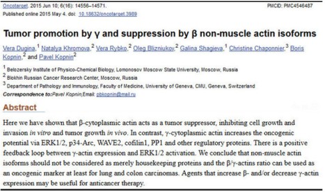
It didn’t go unnoticed by me that this paper failed to cite any of our papers, which had produced fundamental knowledge about human cytoplasmic actins. For example, instead of citing our 1980 paper on the amino acid sequences of human cytoplasmic beta- and gamma-actins, the Russian authors cited a paper on the sequences of bovine actins. Furthermore, these authors were apparently unaware of our discovery of actin mutations leading to tumorigenesis and several examples of null alleles of human beta-actin genes associated with tumors.

I communicated by email with the senior author of this paper, Pavel Kopnin at the Blokhin Russian Cancer Research Center in Moscow, not to complain about these omissions, but to tell him that I liked his hypothesis and to explain why. He thanked me and opined that he had had trouble persuading reviewers to publish the paper. I told him that our findings supported his hypothesis and would have made his argument stronger. He apologized for not citing our work and said that he had not reviewed the literature that far back, which amounted to twenty-eight years since our last paper from LPISM was published in 1987 (this made me feel old).

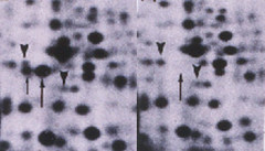
As early as March 1980, I had suggested in writing that altering the ratio of beta- and gamma-actins might contribute to the causation of cancer. This paper was published in the major journal, Journal of Biological Chemistry (see the figure below, last sentence of the abstract). If Dugina et al. were to consider filing a patent on this idea as an invention, our paper would have to, at least, be considered as invalidating prior art along with the rest of our work at LPISM up to 1987.

andy

Both our work at LPISM and Schoenenberger’s work in Basel indicate that the mutation in one of two alleles of the beta-actin gene produces a stable, but defective, form of beta-actin. If Dugina’s hypothesis is correct, it is tempting to suggest that the function of the mutation site in beta-actin controls suppression of tumor formation. I recommended to Pavel Kopnin that his lab pursue this and it is my impression that his lab will continue to work on this hypothesis.

In our model system, we isolated a derivative cell line from the original mutated human fibrosarcoma cell line that exhibited even faster tumor formation (Leavitt et al, 1982). In this second cell line, the mutant beta-actin gene had acquired two additional mutations that made the mutant beta-actin labile with a fast turnover rate in the cell (Lin et al, 1985). As the result of this change, the ratio of stable beta- to gamma-actin changed from approximately 2:1 to approximately 1:1. Furthermore, we found that the two remaining stable forms of beta- and gamma-actin up-regulated in synthesis to maintain a constant normal amount of actin in the cell.

In addition, when we transferred additional mutant human beta-actin genes into immortalized but non-tumorigenic human fibrosarcoma cells, we found that both beta- and gamma-actin from the endogenous normal genes were down-regulated to maintain a constant stable amount of actin in the cell. Thus, we found and reported that beta- and gamma-actin levels in living cells auto-regulated the activities of their own endogenous genes to maintain a constant level of actin in the cell along with a constant ratio of these actins as well (Leavitt et al, 1987a; and Leavitt et al, 1987b). This finding was later confirmed by other laboratories.

These final observations lend support to the idea that maintaining a normal ratio of fully functional cytoplasmic beta- and gamma-actins may be required for the maintenance of the normal, non-neoplastic cellular phenotype. By contrast, mutations and deletions that alter the ratio of functional cytoplasmic beta-actin to gamma actin could lead to tumorigenesis. Hopefully, Pavel Kopnin and others who are aware of our work at LPISM will explore further the role of cytoplasmic actins in maintenance of the normal, non-neoplastic state.

L-Plastin is One of 70 Signature Genes Used to Predict Prognosis of Breast Cancer Metastasis

[Guest post written by John Leavitt, Ph.D., retired Senior Scientist at LPISM in Palo Alto CA from 1981 to 1988; living in Woodstock, CT.  Leavitt has contributed several posts to the Pauling Blog in the past, all of which are collected here.]

John

John Leavitt

On August 24, 2016, the New York Times summarized the results of a Phase 3 clinical study of 6693 women with breast cancer. The outcome of this extensive clinical study was published in the New England Journal of Medicine on August 25, 2016. The clinical trial had been initiated ten years earlier on December 11, 2006 in Europe, (2005-002625-31) and on February 8, 2007 in the United States (NCT00433589). The study examined seventy select genes (seventy breast cancer “signature genes”) out of approximately 25,000 genes in the human genome that, when assayed *together* using a high density DNA microarray, predict the need for early chemotherapy.

In other words, the study asked which of the 6,693 tumors were “high risk” and likely to metastasize to distant sites within a five-year period, and which of these tumors were “low risk” and likely not to metastasize to distant sites in five years. One stated purpose of the study was to determine the need for chemotherapy, which can be very toxic and cause unnecessary harm to the patient, in treating breast cancer. The study found that a certain pattern of elevated or diminished expression of the seventy signature genes can predict a favorable non-metastatic outcome without chemotherapy for five years (while undergoing other forms of therapy such as surgery and irradiation).

One of the seventy selected genes is L-plastin (gene symbol “LCP1” and identified by the blue arrow in the figure below).

List of 70 signature genes

In 1985, my colleagues and I identified this protein in a cancer model system and named it “plastin” (Goldstein et al., 1985). We cloned the gene for human plastin while at the Linus Pauling Institute of Science and Medicine in 1987, and discovered that there were two distinct isoforms encoded by separate genes, L- and T-plastin (Lin et al, 1988). In 2014, in a piece published on the Pauling Blog, I described in some detail the discovery of L-plastin and its subsequent cloning.

A second figure, which is included below, summarizes information about L-plastin in a gene card published by the National Center for Biotechnology Information. This card shows that “LCP1: is the gene symbol for L-plastin and also identifies alternative names for L-plastin. Except for the inappropriate expression of L-plastin in tumor cells, this gene is only constitutively active in white blood cells (hematopoietic cells of the circulatory system). We used very sensitive techniques to try and detect L-plastin in non-blood cells such as fibroblasts, epithelial cells, melanocytes, and endothelial cells, but could not detect its presence in these normal non-hematopoietic cells of solid tissues.

Plastin Gene Card

The L-plastin gene card.

The clinical study reported on in the New York Times and New England Journal of Medicine shows that if L-plastin is not elevated in synthesis and modulated in combination with other signature genes, there should be little or no metastasis in five years. However, if L-plastin, in combination with other signature genes, is elevated in the early stage tumor, then the tumor is a high risk for metastasis and should be treated with chemotherapy.

plastin gels

The above figure consists of a pair of two-dimensional protein profiles that show the difference in expression of L-plastin and its phosphorylated form (upward arrows) between a human fibrosarcoma (left panel) and a normal human fibroblast (right panel).

My colleagues and I also found that L-plastin elevation is likewise a good marker for other female reproductive tumors like ovarian carcinoma, uterine lieomyosarcoma and choriocarcinoma (uterine/placental tumor), as well as fibrosarcomas, melanomas, and colon carcinomas. Abundant induction of L-plastin synthesis was likewise observed following in vitro neoplastic transformation of normal human fibroblasts by the oncogenic simian virus, SV40 (see Table IV in Lin et al, 1993).

The abundant synthesis of L-plastin that we found normally in white blood cells (lymphocytes, macrophages, neutrophils, etc.) suggested to me that the presence of L-plastin in epithelial tumor cells like breast cancer cells contributes to the spread of these tumor cells through the circulatory system to allow metastasis at distant sites. Indeed, both plastin isoforms have now been linked to the spread of tumors by metastasis, an understanding that is summarized in another Pauling Blog article from 2014 and, more recently, in other studies.

The Discovery of Human Plastin at the Pauling Institute

Milestones in Plastin Research

[Guest post written by John Leavitt, Ph.D., Nerac, Inc., Tolland, CT.]

In 1985 my lab at the Linus Pauling Institute of Science and Medicine (LPISM) in Palo Alto, California started to work on an abundant protein of white blood cells (lymphocytes, macrophages, etc.) that mysteriously appeared in human tumor-derived cells of solid tissues (carcinomas, fibrosarcomas, melanomas, etc). I had noticed this phenomenon a few years earlier while at the National Institutes of Health. I also noticed that this protein appeared in oncogenic virus-transformed (SV40 virus) human fibroblasts, but the protein was not expressed in the normal fibrolasts.

I was intrigued by the fact that a major protein of circulating blood cells would be induced during solid tumor cell development because it is well known that solid tumor cells become more anchorage-independent and can circulate like white blood cells to metastasize to other organs. My colleague, David Goldstein, took the lead in examining the expression of this mysterious protein in different cell types of fractionated white blood cells. At the time this protein was assigned only a number (p219/p220) corresponding to its position in two-dimensional protein profiles. We found that this protein was abundantly expressed in all normal white blood cell types that we examined but it was not expressed in normal cells of solid tissues (Goldstein et al, 1985).

When David’s paper was submitted to Cancer Research, the reviews came back positive and the paper was accepted for publication, but one reviewer asked that we give the protein a name. I was thrilled by the thought of naming a protein and its gene which would immortalize our work, so I took on the serious task of coming up with a name that had lasting meaning. My theory was that this cancer marker contributed in some then-unknown way to the plasticity of the cytoplasm in solid tumor cells because of its normal presence in circulating white blood cells. Also, I had seen the great movie, The Graduate, with Dustin Hoffman and recalled that amusing scene depicted in the picture included below. So I named the protein “plastin” – the greatest new thing since sliced bread. 🙂

The Graduate

That same year, I met Steve Kent from Caltech at a meeting in Heidelberg, Germany. After hearing my talk, Steve suggested that we collaborate. He mentioned that a postdoctoral fellow in Leroy Hood’s lab, Dr. Ruedi Aebersold, was trying to develop a more sensitive protein sequencing method for purposes of determining snippets of amino acid sequences from small amounts of unknown proteins eluted from two-dimensional gels (protein profiles) like the gels that we used to characterize plastin in David’s paper. If we could get an accurate partial sequence of plastin, we could devise a nucleic acid probe based on the genetic code that could be used to clone a plastin “copy DNA” from a cDNA library. If the plastin cDNA was cloned, we could then define the protein and perhaps its function by determining the nucleic acid coding sequence in the clone.

Madhu Varma.

Madhu Varma.

I gave Dr. Madhu Varma at LPISM the arduous task of isolating the plastin polypeptide “spot” for sequencing. Madhu cut out the stained spot from 140 two-dimensional gels, in effect purifying enough protein for sequencing by Ruedi at Caltech. Madhu succeeded and Ruedi produced eight short peptide sequences that could be used to develop short nucleic acid probes that would hybridize to the plastin cDNA clone isolated from a tumorigenic human fibroblast cDNA library.

Ching Lin.

Ching Lin.

Dr. Ching Lin at LPISM took one of the nucleic acid probes and immediately attempted to screen a tumorigenic fibroblast cDNA library. If we identified any clones that bound this probe, then we would perform a quick test to determine that we had cloned the plastin coding sequence. But science is full of surprises and we found that the first clone he isolated detected a gene product that was not in lymphocytes but only in normal human fibroblasts – in other words, it failed the test. This is where Ching’s brilliance took over. He was convinced that this first clone he had isolated was indeed a plastin coding sequence so he used this clonal DNA as a new probe against the tumorigenic fibroblast cDNA library. He isolated a new clone that passed the test and detected a gene that was expressed in lymphocytes and tumorigenic fibroblasts but not in normal human fibroblasts.

We performed other experiments that proved that we had cloned two different isoforms of plastin: L-plastin, expressed in lymphocytes and solid tumor-derived cells, and T-plastin that was expressed in normal solid tissues and co-expressed with L-plastin in tumor cells from solid tissues (Lin et al, 1988; Lin et al, 1990). Ultimately this work led to the complete characterization of the human plastin multigene family and verification that both isoforms were aberrantly expressed in various types of human tumors.

The figure at the top of this post maps the progression of discovery that followed our research, which began at the Pauling Institute in 1985. Our publications are shown in red in the graph and research published by other labs is shown in the blue bars.

Here are several plastin milestones discovered by other researchers:

  • T-plastin is abundantly induced in Sezary lymphomas, a lethal T-lymphocyte cancer (Su et al, 2003);
  • L-plastin induction in solid tumors contributes to invasive cancer growth and metastasis (Klemke et al, 2007);
  • Mutations in T-plastin play a role in the genetic disease Spinal Muscular Atrophy (Oprea et al, 2008); and
  • Most recently mutations in both L- and T-plastin promote re-growth of colon carcinomas following surgical resection of these tumors and chemotherapy (Ning et al, 2014).

These developments are more or less typical of the way science works. Progress in understanding complex phenomena like human cancer is the work of many scientists that builds on the observations of other scientists. This is just one example of the productive contributions in biomedical research that came about through early discovery research at LPISM in the 1980s.

Research Completed at LPISM in 1988 – Reproduced and Extended in 2014

The author in his laboratory at the Linus Pauling Institute of Science and Medicine. Originally published in Science Digest, June 1986.

The author in his laboratory at the Linus Pauling Institute of Science and Medicine. Originally published in Science Digest, June 1986.

[Guest post written by John Leavitt, Ph.D., Nerac, Inc., Tolland, CT.]

In 1987, my colleagues at the Pauling Institute in Palo Alto, colleagues at Stanford and I published a paper that clearly demonstrated that expression of a charge-altered mutant human beta-actin (glycine to aspartic acid substitution at amino acid 245; G245D) caused non-tumorigenic, immortalized human fibroblasts to form aggressive tumors in nude mice (Leavitt et al, 1987a). When these tumor-derived cells were examined, we discovered that they exhibited further elevated expression of the mutant beta-actin and these tumor-derived cells formed tumors even more rapidly – observations that were consistent with the role of this mutation in the tumorigenic phenotype. Furthermore, over-expression of mutant beta-actin was associated with down-regulation of three abundant tropomyosin isoforms in a well-documented transformation-sensitive manner (Leavitt et al, 1986; Leavitt et al, 1987a and Ng et al, 1988). These final papers were the culmination of research conducted at the Linus Pauling Institute of Science and Medicine (LPISM) from December 1981 to March 1988.

Normally when a scientific observation is never repeated it is usually not worth remembering. In this case, twenty-six years after our 1987 publication, a study was published by Schoenenberger et al. at the Biozentrum in Basel, Switzerland, that reproduced our findings in a different cell system, a rat fibroblast model (provided to them by LPISM in 1986). Furthermore, these investigators extended our findings by characterizing new aspects of abnormal behavior of the mutant beta-actin and cells that express this aberrant protein, which help to explain its potential role in cancer such as enhancement of tumor cell motility and invasiveness.

In addition to enhancement of tumor growth and alteration of cell shape, the Swiss investigators presented the following findings to clarify and support the oncogenicity of this mutation:

  1. The mutant actin stimulated formation of ruffles at the cell periphery as shown by staining of cells with an antibody that bound specifically to the mutant epitope of the mutant beta-actin (left image below)
  2. The mutant actin concentrated primarily in these ruffles (palloidin staining reveals the location of filamentous actin in stress fibers; right image below)
  3. The expression of mutant actin inhibited the tropomysin binding to filamentous actin and tropomysin did not accumulate in the ruffles
  4. Mutant actin colocalized with Rac1 (a GTPase mediator of membrane ruffling) and beta1-integrin (adhesion protein) in the ruffles

ruffles

Back-tracking several years, the discovery of this actin mutation was made in a mutagenized cell line isolated by Takeo Kakunaga at the National Cancer Institute (NCI) in 1978. During the month that his paper was published, I walked over to NCI from my lab across the street at the Bureau of Biologics (FDA) to have a chat with Takeo about using his in vitro transformed Syrian Hamster cells as a model system to identify changes in protein expression that correlated with neoplastic transformation. After describing what I wanted to do, he seemed agreeable but then casually mentioned that he had succeed in transforming human fibroblasts into tumor forming cells. I nearly fell off my chair because human cells had never been transformed in vitro before, a major problem for cancer researchers at that time.

I blurted out that we should do the work that I had proposed in his human cell model system, comparing protein expression by the transformed neoplastic cells with their normal precursor cells. My hypothesis was that this comparison would allow identification of proteins that were turned on or turned off in expression by comparing protein profiles of the most abundant 1,000 proteins expressed in these cells and resolved by high-resolution 2-D gel separation (protein profiling). My plan was to look for charge-altering mutations in proteins that might govern neoplastic transformation and tumorigenesis. A fall-back goal was to define the pattern of qualitative and quantitative changes in protein synthesis to try and get a handle on the mysterious mechanism of human cancer development. A summary of the global changes in gene expression of neoplastic human fibroblasts was published from LPISM in 1982 (Leavitt et al, 1982).

Within two weeks, in May of 1978, I was metabolically labeling the total cellular proteins (with the amino acid S-35 methionine) of the normal fibroblasts and three strains of cell lines derived from the normal culture which were immortalized, only one of which formed subcutaneous tumors in nude mice. After four hours of labeling, I prepared extracts of S-35 methionine labeled proteins from each of the four cultures and loaded 25-microliter aliquots of each sample onto the top of clear noodle-like isoelectric focusing gels (7-inch long urea-polyacrylamide gels with the thickness of thin spaghetti) which separated the complex mixture of total cellular proteins by their net charge (isoelectric point). These gels were subjected to isoelectric electrofocusing of the proteins overnight. The next morning I harvested the spaghetti-like gels, and incubated them in a detergent that would bind to the proteins to help separate them by their molecular weights in a second dimension. So, these proteins were first denatured and separated by their net charge and then, in a second dimension, separated by their size on a thin rectangular slab gel.

After about five hours of separation in the second dimension, I was soon to learn that I had separated more than 1,000 denatured protein subunits (polypeptides) by their differing charges and molecular weights. The final step before autoradiography, which revealed the full protein profile, was to fix and stain the gels to get a glimpse of the resolution of these peptide patterns. The staining of these rectangular gels revealed only the most abundant architectural cellular proteins, the largest number of which were cytoplasmic beta- and gamma-actin, at a ratio of about 2:1 in abundance, respectively. The figure below shows what quickly appeared as the gels were de-stained. In the one tumorigenic cell line, instead of seeing a 2:1 ratio of beta- to gamma-actin, a new abundant protein at about one unit charge more negatively charged (more acidic), and half of the normal beta-actin was lost. The pixilation of these three radioactive “spots” immediately suggested to me that one of the two functional genes (alleles) encoding beta-actin had mutated, possibly due to the replacement of a neutral amino acid with a negatively charged amino acid. This prediction was no mystery to me as I had demonstrated this type of electrophoretic shift in another protein a year earlier at Johns Hopkins.

mutant actin further annotated

A number of experiments were done to build the case for the beta-actin mutation, and then I wrote a letter to Klaus Weber at the Max-Planck Institute in Goettingen, Germany, asking for his help in sequencing these actins. His lab was the only one in the world sequencing actins, e.g. the four muscle forms of actins. It only took Klaus two weeks to respond affirmatively, an indication to me that he was eager. I provided him with the actin proteins from this cell line and it took a postdoctoral fellow, Joel Vandekerhkove, and Klaus a little over a year to determine the complete amino acid sequences of the mutant beta-actin and both the wildtype beta- and gamma-actins, to define the mutation that had occurred. We published the result shown above in the top journal Cell in December 1980. Four years later, my colleagues at Stanford and I published my cloning of the mutant and wildtype human beta-actin gene, and the experiments that formally proved the mutation at the level of the gene (Leavitt et al, 1984). Three years after that, we published the experiments that demonstrated the tumorigenic effect of this mutation in immortalized human fibroblasts.

The dramatic nature of this discovery was never fully appreciated, perhaps, because no other actin mutations had been reported and it took Scheonenberger, et al. twenty-six years to complete the work published in September 2013. In another recent related development, Lohr et al. reported reoccurring beta-actin mutations in a panel of tumor cell samples from patients with diffuse large B-cell lymphoma.

One interesting piece of information that came out of our initial sequencing of these actins was the degree of evolutionary conservation of human beta- and gamma-actin. These two actins differ by only four amino acids at the N-terminus, whereas the four muscle-specific human isoforms are more divergent. Comparing the sequence of actin cloned from Saccharomyces cerevisiae (yeast) with these human sequences (sequences stored at the National Center for Biotechnology Information; NCBI) reveals that yeast and human cytoplasmic actins are 92% identical in their sequences (differing by only 31 amino acids out of 375) and most of these amino acid exchanges are conservative replacements both structurally and thermodynamically. This makes these actins the most highly conserved proteins (on a par with histones H3 and H4) among the 20,000 or so known human protein sequences. This fact presents an argument for the fundamental importance of non-muscle cytoplasmic actins in eukaryotic life. It turns out that among actin sequences of all species, no replacement of the Glycine 245 has ever been documented as a result of species divergence or mammalian isoform divergence.

When we introduced the mutant beta-actin gene into a non-tumorigenic immortalized fibroblast strain by gene transfer (Leavitt et al, 1987a), we isolated a transfected clone in which the ratio of exogenous mutant beta-actin to wildtype beta- + gamma-actin was 0.88 – a 76% higher level of expression than the mutant actin in the original mutated cell line in which the mutation arose (0.5 ratio). When we isolated and cultured the cells from a tumor formed by this cell line, the ratio of exogenous mutant beta-actin to wildtype beta- + gamma-actin had increased to 1.95, indicating that about 64% of the total cytoplasmic actin was the mutated beta-actin. Whereas the initial transfectant cell line produced visible tumors at about six weeks, the tumor-derived transfectant cells expressing 64% mutant actin formed visible tumors at about 1.5 weeks. Thus, expression of this mutation was not inhibitory to cell growth.

The other surprising finding was that cell lines expressing the transfected mutant actin gene did not have higher levels of cytoplasmic actins in them because the two endogenous wildtype beta- and gamma-actin genes were coordinately down-regulated (auto-regulated) so that the relative rates of total actin synthesis remained around 30% compared to S-35 methionine incorporation into 600 surrounding non-actin polypeptides in the protein profile (Leavitt et al, 1987b). This auto-regulation phenomenon was reproduced by Minamide et al. (1997) ten years later.

Cytoskeletal rearrangement of actin microfilaments, as well as changes in composition of tropomyosin isoforms and other actin-binding proteins, have long been associated with neoplastic transformation. However, before our study, causal mutations in a cytoplasmic actin had apparently not been considered. It is perhaps consistent then that Ning et al. (2014) have recently described genetically inherited polymorphisms in the actin-bundling protein, plastin (also discovered and cloned at LPISM), that significantly affect the time of tumor recurrence in colorectal cancer after resection and chemotherapy.

During my tenure at the Pauling Institute, I felt that Dr. Pauling understood and appreciated this work and its relevance to the fundamental nature of cancer development. Progress can be slow, but ultimately true understanding of cancer will emerge from this type of research…and I predict that cytoplasmic actins and actin-binding proteins that regulate actin organization and function in the cytoskeleton will be understood to play a central role in the manifestation of the tumorigenic phenotype.

Some Personal Thoughts on Vitamin C in the 1980s and Now

[Guest post written by John Leavitt, Ph.D., Nerac, Inc., Tolland, CT.]

The author in his laboratory at the Linus Pauling Institute of Science and Medicine. Originally published in Science Digest, June 1986.

The author in his laboratory at the Linus Pauling Institute of Science and Medicine. Originally published in Science Digest, June 1986.

During my daily work for pharmaceutical and biotech clients, I am continuously learning about developments resulting from my research at the Linus Pauling Institute of Science and Medicine in Palo Alto, CA in the 1980s. Likewise, I am regularly coming into contact with new medically related developments focusing on vitamin C, an interest of Linus Pauling in those years.

With regard to our research on human plastins, a gene family of proteins that we discovered, cloned, and characterized at the Pauling Institute, it has recently been reported that plastin (PLS3) is a marker of carcinoma cells circulating in the blood (for example Yokobori, et al.). Our hypothesis was that when this protein was inappropriately expressed in cells from solid tissues, as it is in many tumor types, (e.g. carcinomas, fibrosarcomas, etc.) these potential tumor cells become more like blood cells in that they are able to live and replicate in an anchorage-independent state, an essential property of metastatic tumor cells. It is metastasis that kills us when we get cancer. Thus plastins, discovered and characterized at the Pauling Institute, may turn out to be the “holy grail” of cancer research.

I often run across new information on the medical importance of vitamin C without looking for it. Back in the 1980s, we would receive an annual shipment of loose vitamin C from Hoffmann-La Roche, Inc. as a way of saying thank you to Dr. Pauling for his advocacy of the merits of vitamin C. We received no funding from Hoffmann-La Roche though. One year I recall that two dignitaries from the company visited us. Dr. Pauling, with me and several others, walked our visitors to lunch a few blocks down El Camino Real in Palo Alto to my favorite restaurant, the Captain’s Cabin.

Afterward, while walking back to the Institute, one of the guests asked Dr. Pauling if he thought the perceived benefits of vitamin C were due to the placebo effect. I was amused because I too had said something ill-advised like that to Dr. Pauling in my first few months at the Institute. I mentioned to him that I had a vitamin C-resistant cold to which he replied, “You’re not taking enough!” and told me that he takes 18 grams a day. No doubt he had calculated this number based on the amount of vitamin C that animals produce within themselves every day. He would stir 18 grams into a large glass of water and imbibe the glass with no great rush.


A few months ago I heard a physician state in the national media that taking supplemental vitamins is a waste of money. This bold assertion reminded me of the announcement of the discovery of cold fusion and another premature announcement of the discovery of a cure for AIDS. The progress of science is slow but relentless, like the new developments with plastins fifteen years after I left LPISM’s labs.

On October 31, 2013, Kim, et al. at Seoul National University in South Korea published their findings on a new strain of experimental mice. The researchers knocked out the mouse gene encoding the enzyme L-gulono-γ-lactone oxidase, known as gulo for short. This is the gene that is missing in humans and that keeps us from synthesizing our own vitamin C, unlike nearly all other animals. An extreme lack of vitamin C in our diet can lead to scurvy, caused by aberrant expression of collagen in our connective tissues because of starvation of vitamin C in our diet. In these mice the lack of this gene caused “vitamin C insufficiency” in an animal model – a model that can now be used to learn more about the importance of vitamin C.

As these mice matured they expressed known blood markers of liver damage. This damage, called fibrosis, is basically the scarring of the liver, sort of like the scarring of the skin that is caused by certain types of skin damage. Concomitantly, as the mice aged, reactive oxygen species (ROS) and lipid peroxides increased in the liver, as did activated hepatic stellate cells, which deposited abnormal collagen fibriles on the basement membrane of functional liver cells. There is a wealth of evidence that elevated ROS in the lungs, liver, and kidneys is associated with pulmonary, hepatic, and renal fibrosis. Elevated vitamin C in these tissues will quench ROS.

Currently in the United States, there are no drugs approved to treat any of these forms of fibrosis. Fortunately, Intermune’s drug, pirfenidone, is close to approval for treatment of pulmonary fibrosis and has already been approved in Canada, Europe, and Japan. This drug reduces ROS and inhibits other key targets that are suspected of playing a role in the development of fibrosis. So who is to say that supplementing your diet with vitamin C is of no consequence? It is certainly not toxic in any way. Oh, by the way, pulmonary fibrosis is worse than cancer – it kills you in three to five years once diagnosed. You basically die of asphyxiation.


In the last week I stumbled upon another interesting paper on the effects of vitamin C on humans. A 2011 paper by Juraschek, et al. at Johns Hopkins University Medical School reported the results of a significant meta-analysis (a systematic review of multiple clinical trials) of 13 randomized clinical trials involving 556 patients who took a median dose of 500 mg of vitamin C per day. (I take a full gram)

The purpose of the study was to examine the effects of vitamin C supplementation on uric acid levels in the blood. Elevated uric acid levels in the blood causes gout, because saturation of blood with uric acid causes urate crystals to form in the synovial fluids of joints (e.g. crystal arthritis). Drugs that lower uric acid in the blood are used to treat gout because lowering uric acid causes the urate crystals to dissolve to ameliorate the arthritic pain.

Admittedly gout is not as bad as cancer. But another systematic clinical review of multiple trials on humans published in 2012 by Lottmann, et al. at the IGES Institut GmBH in Germany has shown clearly that having gout is associated with both all-cause mortality and, in particular, cardiovascular mortality. So what could be worse than death by gout?

I think I will keep taking vitamin C.

Dr. Pauling’s Prediction of a Mutation in Beta-Globin Which Causes Sickle Cell Anemia and How This Prediction Impacted My Research

[Guest post written by John Leavitt, Ph.D., Nerac, Inc., Tolland, CT.]

Linus Pauling lecturing on sickle cell anemia, Kyoto, Japan. 1955

In September 2010, the company BlueBird Bio announced that it had cured a patient with the hemoglobinopathy, beta-thalessemia, by correcting the genetic defect in beta-globin that this patient inherited from his parents. This came 61 years after Linus Pauling and his associate, Harvey Itano, explained the cause of hemoglobinopathies such as sickle cell anemia. Beta-thalessemia like sickle cell anemia is caused by an inherited mutation in the beta-globin gene, just a different mutation. In the case of thalessemia, the defective beta-globin gene product disappears, whereas the defective beta-globin in sickle cell anemia remains stable to wreak havoc on the body. BlueBird Bio accomplished this first cure of a hemoglobinopathy by removing the blood-forming hematopoietic stem cells from the patient, engineering his cells ex vivo with a correct beta-globin gene, and then putting the cells back into the patient. The stem cell transplant sustained itself and produced red blood cells which functioned normally in the circulatory system. For the first time in this patient’s 18 year-old life, he did not have to have a monthly blood transfusion.


In late September 1981, when I gave a seminar at the Pauling Institute of Science and Medicine in Palo Alto, CA, I noticed that Dr. Pauling was smiling during the talk. He was aware of the discovery of the muscle isoform of actin by his friend Albert Szent-Györgyi and knew about the structure and function of actins (the subject of my talk). After reading Dr. Pauling’s 1949 paper on the molecular nature of the sickle cell trait, I understood that he was seeing during my talk the very same experiments in my discovery of a mutant human beta-actin that he and Harvey Itano had performed, which led to the prediction of a mutation in the hemoglobin protein that caused sickle cell anemia. His paper was the very first to describe the molecular genetic basis of a human disease. By 1981 there was plenty of conceptual evidence to suggest how I could look for mutations in proteins using electrophoretic separation of complex mixtures of cellular proteins. In 1949 though, Dr. Pauling was way ahead of his time. In his and Itano’s case, the plan was well thought out based upon years of characterization of oxygen bonding to the heme of the globin molecule. By contrast, I was very lucky to find a mutation in the most abundant structural protein of the cell, cytoskeletal actin in a human fibrosarcoma cell.

Harvey Itano.

It was probably evident in 1949 that hemoglobin amounts to about 95 percent of the total protein of a mature red blood cell; so these cells were essentially bags of hemoglobin molecules – globin polypeptides with attached heme moieties with an iron atom that bound oxygen. The heme-bound iron carries oxygen through the arterial system to cells for respiration. After delivery of oxygen to tissues, these red blood cells (RBCs) return carbon dioxide to the lungs through the venous system for expiration. In sickle cell anemia, after RBCs deliver oxygen throughout the body, the RBCs take on a sickled shape, clog the venous system and lyse, causing a wide variety of systemic problems. Pauling and Itano predicted that this change in RBC architecture was a direct consequence of “two to four” charged amino acid changes in the globin complex, which consists of two beta-globin subunits and two alpha-globin subunits (this was not known then). Because of the science that came after their discovery, we know now that the genetic mutation in the beta-globin moiety is a single amino acid exchange of glutamic acid to valine resulting from a single nucleotide transition (A to T transition) in codon 6 of the beta-globin gene encoding the 147 amino acid polypeptide. Thus two positive charges were added to the hemoglobin molecule by this mutation. Pauling and Itano concluded that these charge alterations caused RBC sickling.

Important discoveries can be quite simple. The figure below is the key experiment carried out by Pauling and Itano, an electrophoretic separation of hemoglobin based upon its isoelectric point (net charge). Because of the mutation in codon 6 present in both inherited beta-globin alleles, the hemoglobin complex migrated to the right of the normal hemoglobin by approximately “two to four” positive charges (panel B compared with panel A). At pH 6.9 the normal hemoglobin was shown to have an isoelectric point of 6.87, migrating as a negative ion, whereas the mutated hemoglobin had an isoelectric point of 7.09 migrating as a positive ion. We know now that this electrophoretic change in the hemoglobin complex described by Pauling and Itano is due to the loss of a single negative charge in a glutamic acid residue (replaced with an uncharged valine residue) near the N-terminus of the two beta-globin moieties of the hemoglobin molecule. Today, the fact remains that this is the only mutation in hemoglobin that causes sickle cell anemia, although other beta-globin mutations cause other hemoglobinopathies like beta-thalessemia. Panel C shows the electrophoretic behavior of hemoglobins in a heterozygous carrier of the disease-causing mutation (Panel D is a control mixture of the globins in panels A and B). Much more insight about these phenomena is discussed in the Pauling and Itano paper but the charge alteration in hemoglobins is the basic observation.

Pauling experiment

(click to enlarge)

Fast-forward to 1976. I decided to look for evidence of charge-altering mutations in a protein profile of about 1,000 visible proteins (polypeptides) comparing normal and neoplastic cells by looking for Pauling and Itano’s evidence of mutations, e.g. minor charge alterations in proteins in the protein profile. A technique had just been developed by Patrick O’Farrell which permitted high-resolution separation of virtually all major protein gene products of the cell.

An elegant study was performed by Greg Milman at the University of California at Berkeley who demonstrated that one could predict the occurrence of mutations in the relatively minor protein, the enzyme hypoxanthine phosphoribosyltransferase (HPRT), in HeLa cells by the positional changes in the HPRT polypeptide in high-resolution two-dimensional polyacrylamide gels within complex profile of proteins separated both by their charge (isoelectric point) and their molecular weight. When I saw Milman’s result I decided to use this technique to compare normal and neoplastic human cells to see if I could identify charge alterations similar to those demonstrated by Pauling and Itano in hemoglobin and by Milman in HPRT.

I labeled the proteins of normal and tumor-forming human fibroblasts with S35-methionine and separated them using O’Farrell’s two-dimensional technique (isoelectric point separation is a tube gel followed by molecular weight sieving in an SDS slab gel). Then I fixed the proteins in the two-dimensional slab gel and stained these proteins with Coomassie blue dye.

mutant actin further annotated

With the dye you could only see the most abundant proteins and I was surprised to see this pattern of actins in the tumor-forming fibroblasts shown above. This image is actually the image of the radioactive protein pattern in the region of actins (pI 5.3 to pI 5.1, molecular weight Mr about 42,000) developed after a very short autoradiographic exposure to X-ray film (a digital image). Normally you only see one beta-actin spot barely separated from the gamma-actin spot. Gamma actin is a second isoform of actin encoded by a separate gene which differs by only four amino acids from beta-actin. Normally there is about twice as much beta-actin at isoelectric point 5.2 as gamma actin and both actins together amount to 5-10 percent of the total cellular protein. But half of the normal beta-actin was missing and a new more negative spot at isoelectric point 5.3 appeared. I was able to show that this was a new form of beta-actin by tryptic peptide separation and other criteria. The observation that the new variant migrated slower in the second dimension as a larger protein was later attributed to a frictional effect in the gel sieve due to an altered conformation caused by the amino acid change.

These observations and other differences in protein expression between the normal and tumor-forming fibroblasts were published in the Journal of Biological Chemistry in February 1980. A second paper was published a month later demonstrating that a T-cell leukemia also had a beta-actin anomaly which suggested loss of a beta-actin allele. It is now well established that reorganization of the actin cytoskeleton occurs when cells become cancerous, although mutations in the structural gene may be less common. These alterations can also be caused by changes in actin-binding proteins.

Later in the year, with my colleagues at the Max-Planck in Goettingen Germany, Klaus Weber and Joel Vandekerckhove, I published the sequences of the normal human beta- and gamma-actins and the mutant beta-actin in Cell. The normal and mutated sequence of human beta-actin is shown in the figure below.

The simple electrophoretic difference between the mutant and normal beta-actin was a single amino acid exchange of a neutral glycine for a negatively charged aspartic acid at amino acid residue 244 in the 374 amino acid polypeptide chain, an observation similar to Pauling and Itano’s hypothesis 32 years earlier. An amino acid exchange at this residue in the actin polypeptide chain had never been observed in any eukaryote. Two years later I cloned the mutant and wildtype human beta-actin genes at the Pauling Institute and formally proved the existence of the mutation at the level of the gene. This mutation was caused by a single nucleotide change in the gene. Several years later my colleagues and I demonstrated that acquisition of this simple mutation contributed to the tumorigenic phenotype of the cells in which it arose.

actin sequence with arrow

The sequence of human beta-actin and its amino acid 244 mutation (the most highly conserved protein in eukaryotes).


Ed Note: This week marks the sixth anniversary of the creation of the Pauling Blog.  Birthed to help promote the unveiling of a postage stamp, the blog, 461 posts later, has developed into a resource of consequence with an audience that is steadily growing.  For those who might be interested in how the project operates, please see this post that we ran one year ago.

As always, we thank you for your continued readership.  We plan to keep researching and writing, so please keep coming back!

1955i.45

Pioneering the Field of Proteomics

John Leavitt, 1982.

John Leavitt, 1982.

[Guest post written by John Leavitt, Ph.D., Nerac, Inc., Tolland, CT.]

In the fall of 1985, I went to a small meeting in Heidelberg, Germany, with Steve Burbeck from the Linus Pauling Institute of Science and Medicine, who had helped me by developing computerized microdensitometry to analyze two-dimensional protein profiles. At this meeting I described our protein profiling work and the discovery of the mutant beta-actins and another interesting protein which I named “plastin.”

Steve Kent, head of the protein sequencing facility in Leroy Hood’s lab at Caltech, heard my talk. We sat across from each other at dinner and he proposed a collaboration to develop methods of sequencing minute amounts of protein leached from spots in high resolution protein profiles. Lee Hood was well known for developing state-of-the-art protein and nucleic acid sequencing methods and machines, and was a founder of Applied Biosystems in Foster City, CA.

After I returned from Heidelberg, Ruedi Aebersold called me from Caltech and we began collaborating on microsequencing of pure nanomolar quantities of unknown proteins of interest eluted out of my protein profiles. In this work we essentially started the field of proteomics, which was eventually named ten years later by Jim Garrells, a protein profiler at Cold Spring Harbor. Proteomics is the search for and definition of proteins that could serve as diagnostic markers and drug targets for diagnosis and treatment of diseases, in our case cancer.

In 1987 we published a landmark paper in PNAS on the microsequencing technique that Ruedi developed. This paper would eventually be cited in references by more than 1,000 other research papers.

Ruedi

I gave a postdoctoral fellow, Mahdu Varma, the task of isolating the cancer-specific leukocyte isoform of plastin (L-plastin) from 140 protein profiles. This protein has been implicated in metastases in both melanoma and prostate cancer as well as in other aspects of cancer. The L-plastin spot was easily recognized and those spots on a nitrocellulose filter were “snipped out,” removing all the other proteins of the cell. We sent Ruedi a plastic tube containing the 140 “spots” of L-plastin. He had figured out a way to solubilize the protein from the nitrocellulose and was successful in determining the sequence of eight oligopeptides of between eight and sixteen amino acids derived by digestion of L-plastin with a proteolytic enzyme.

The peptide sequences he determined turned out to be perfectly accurate internal amino acid sequences of plastin when we decoded the sequence of the plastin gene (cDNA) clone, a reverse transcript of the messenger RNA. This was the first time that anyone had done this and it opened up the field of proteomics and led to the discovery of other diagnostic and drug targets.

plastin 1

We had chosen L-plastin, normally only expressed in white blood cells, because I had reported for years that it was a cancer marker in tumors that arose in solid tissues (identified in the image above by the two upward arrows). After we received the oligopeptide sequences from Ruedi, we made short DNA antisense probes that would hybridize to DNA sequences encoding these peptides in the human genome to fish out the full-length DNA clones that carried the sequence of the L- plastin gene.

Ching Lin and I, along with Reudi, published the sequences of the human L- and newly discovered T-plastin proteins, based upon sequencing of cDNAs, in Molecular and Cellular Biology. The discovery of a second isoform of plastin (T-plastin named for tissue plastin as opposed to L-plastin from leukocytes) was a surprise. We now had two genes to characterize at the genomic level. Today, T-plastin is a well recognized marker for cutaneous T-cell lymphoma (Sezary Lymphoma) and L-plastin, inappropriately expressed in solid tumor cells (carcinomas, fibrosarcomas, etc.), is understood to be a contributor to metastasis.

The Linus Pauling Institute was not all work and no play in the 1980s

We worked hard at the Institute and Linus Pauling was always there and visible.

We put together a softball team with Jim Fleming, Dan McQueeny, Zelek Herman, myself, and others at the Institute and played departmental teams at Stanford. I think we were called the “Pauling squeeze.” After these games we would often go dancing at the Class Reunion on El Camino Real near the corner of Page Mill Road.

We were fortunate to have on staff a first rate fundraiser in Richard (Rick) Hicks who arranged wonderful parties on Nob Hill at the Stanford Court. The most memorable of these parties occurred in late November 1986, when we honored Japanese billionaire Ryoichi Sasakawa with the annual Linus Pauling Medal. Another year Carl Sagan and Ann Druyan, who helped Carl put together the Cosmos series, likewise took part. We often saw Dr. Pauling’s sons, Linus Pauling Jr., Peter, and Crellin as well.

Here we are at the Stanford Court that night with postdoctoral fellows, Dr. Karin Sturm from Heidelberg, Germany, on the left and Dr. Madhu Varma from Madras, India, on the right. My wife, Becki, is in the middle. I recall that Dr. Pauling enjoyed this night as well.

Here we are at the Stanford Court that night with postdoctoral fellows, Dr. Karin Sturm from Heidelberg, Germany, on the left and Dr. Madhu Varma from Madras, India, on the right. My wife, Becki, is in the middle. I recall that Dr. Pauling enjoyed this night as well.

In 1988 I moved on to a new position in San Jose and then became Director of Research at Adeza Biomedical. Since we continued to live in Palo Alto, we continued to interact and party with the Linus Pauling Institute staff into the 1990s.

The 1980s at the Linus Pauling Institute – A Wonderful Place to Be

John Leavitt

John Leavitt

[Ed Note: This is part one of a two part series of guest posts written by John Leavitt, Ph.D., Nerac, Inc., Tolland, CT.]

There was an article about Linus Pauling in Time magazine in early 1981 about the fact that at the age of 80 he was still seeking a grant from the National Institutes of Health (NIH) to fund his research on ascorbic acid for treating diseases. This news caught my attention and I looked into the possibility of joining Dr. Pauling’s institute. Toward the end of the summer I was invited to visit the Pauling Institute in Palo Alto, CA to give a seminar on my research at NIH.

In late August Koloman Laki, an aging scientist at NIH, called me up and invited me over to his lab in NIH Building 10, a short walk across the campus from my lab in NIH Building 37. He was interested in talking to me about my recent discovery of mutations in human non-muscle cytoskeletal actin that was published in Cell in late 1980. This protein is the major architectural protein of all eukaryotic cells and we had shown that it was the most highly conserved protein in evolution of the species from yeast to humans. This fact made these mutations even more interesting.

Koloman was a protege of the Hungarian Nobel Prize winner Albert Szent-Györgyi who, I later learned, was much admired by Dr. Pauling because he had discovered both vitamin C and actin. Koloman described how Szent-Györgyi discovered muscle actin. When I mentioned that I was to visit the Linus Pauling Institute in late September, he told me about Emile Zuckerkandl’s and Dr. Pauling’s work on the ‘biological clock,’ which provided evidence in support of Charles Darwin’s theory on divergence of the species.

In the last week of September I flew to Oakland, CA and was picked up at the airport by Emile who was President of the Linus Pauling Institute of Science and Medicine. The next morning I stood up in front of Dr. Pauling and the institute staff to tell them about my discovery of a mutant human beta-actin and my speculation on its involvement in neoplastic transformation. The evidence suggested that I had actually discovered at least two mutations in the same gene, each of which caused a progression to a higher malignant state.

Linus Pauling was in the front row and was all smiles. He asked me if I knew who discovered actin. I was prepared to answer that question thanks to Koloman Laki. In the afternoon I met with Emile who offered me a Senior Scientist position at the Institute, which I accepted. At the time it would be me and Dr. Pauling with separate research interests. Nevertheless, Dr. Pauling could appreciate my discovery as, 32 years earlier, he had described the molecular basis for sickle cell anemia, which predicted that mutations in hemogloblin governed the sickled shape of red blood cells which caused the disease, sickle cell anemia. Likewise, human cancer cells exhibit altered shapes.

So I resigned my secure job-for-life at NIH and moved to Palo Alto to join the struggling Linus Pauling Institute. My technician, Patti Porecca, hired from Bob Gallo’s lab at NIH, would follow me to the Pauling Institute.

Cloning of the Human Beta-Actin Gene

After I arrived at the Pauling Institute, two of my colleagues at NIH and I published a comprehensive study of the changes in protein expression between normal and neoplastic cells in Carcinogenesis using high-resolution computerized microdensitometry to analyze the complex protein patterns (my first paper from the Pauling Institute). This was the first time that such a study had been published, e.g. the comparative profiling of expression of a large number of proteins in neoplastic cells. It was a study of the 1,000 most abundant proteins in normal and neoplastic human cells which revealed potential biomarkers and causative genetic events for human cancer. At the time it was staggering to view these patterns but perfect for my dyslexic brain and mind’s eye. In addition, we published another paper in Cell that described, for the first time, the progression of a neoplastic human cell to a higher malignant cell following a second mutation in the same beta-actin gene. Early in 1982, Steve Burbeck and Jerry Latter at the Institute set up the same computerized microdensitometry platform I had exploited at NIH.

Jerry Latter gave a stirring talk at Argonne Labs in Chicago demonstrating that computerized microdensitometry of protein profiles could be used to determine the identities of unknown proteins based upon determining their amino acid compositions in situ in protein profiles. This paper was published in Clinical Chemistry in 1984. At the same meeting, Steve Burbeck described a truly innovative invention that could measure beta-particles emitted from radioactive protein profiles to produce a direct image of the protein profile pattern. As a group we had entered an exciting period of discovery and innovation at the Linus Pauling Institute.

When I got to Palo Alto in December 1981, I called Professor Larry Kedes at Stanford and we embarked on a collaboration to clone the human beta-actin gene. His impressive postdoctoral fellow, Peter Gunning, taught me some basic recombinant DNA techniques, and I was off to the races. The difficulty was to identify the functional gene in a sea of actin pseudogenes (sometimes referred to as junk DNA). I used an elegant method of homologous recombination developed in Tom Maniatis’ lab at Harvard that had never been used before to clone a novel gene (In fact, cloning of human genes was just getting started at the time). This was smart because Professor Maniatis would be the chairman of the NIH study section that reviewed my first grant proposal submitted from the Pauling Institute. I did not know it at the time but within a month or two I had cloned the functional beta-actin gene a week before Christmas in 1982.

I developed a scheme to identify the correct gene among 300-400 clones of pseudogenes that Patti and I had cloned and the strategy worked. We gave Dr. Sun-Yu Ng the task of sequencing the DNA clone that we were betting on. Rather quickly we determined that we had cloned the functional human beta-actin gene because the DNA sequence that Sun-Yu determined from our candidate clone accurately encoded the amino acid sequence of human beta-actin protein that I had published in Cell in 1980 (with Klaus Weber). Quite coincidentally another lab discovered a rat oncogene that was a fusion of part of an actin gene with a tyrosine kinase gene. I sent this information off to the study section that was reviewing my grant in January 1984 as added evidence that the actin gene was in some way relevant to neoplasia.

My colleagues and I at the Pauling Institute and Stanford published our successful isolation of both the mutant and wildtype human beta-actin genes in Molecular and Cellular Biology in October 1984. As shown below, we had given Armand Hammer’s name to our cancer research program because of his generosity in helping to fund the Linus Pauling Institute.

actin cloning

In January 1984 I was awarded a grant of about $110,000 a year for two years from the American Cancer Society…what a relief. Later in the spring I received word from Professor Maniatis’ NIH study section that our program would also be funded in June by a grant of about $150,000 a year for 3.5 years from the National Cancer Institute for the same work. I was able to hire Dr. Ching Lin from Iowa State University and Dr. Ng (Sun-Yu) from Kedes’ lab. By 1985 Sun-Yu finished the complete DNA sequencing of the human beta-acid gene and Ching sequenced the copy of the beta-actin gene that had two mutations to formally prove the mutations at the level of the gene. Everything that we had learned about the genetic code and amino acid sequences of proteins made our findings predictable. I had learned from my own research how Darwin’s theory of evolution and natural selection worked.

This was the year I finally successfully transferred in recombinant gene inside a cell in culture. I transferred the mutant human actin gene into a rat fibroblast cell line to show that I had cloned the functional gene which could abundantly express its protein the way the natural endogenous beta-actin gene worked (shown in a protein profile below).

mutant actin annotated

At this point I had a brief meeting arranged by Emile with Alex Zafferoni, founder and CEO of Alza Corporation, a block away on Page Mill Road. Zafferoni recommended Bert Roland as a patent attorney. I arranged a meeting with Roland, also a block away, for that afternoon to discuss patenting the human beta-actin gene promoter because of its strong constitutive nature (the engine of the gene that drives its expression). I told Bert that this was a collaboration with Peter Gunning and Larry Kedes at Stanford. Roland was famous for filing Boyer’s and Cohen’s genetic engineering patent which created Genentech and eventually funded Stanford with hundreds of millions of dollars.

We published Sun-Yu’s work on the sequence, structure, and chromosomal location (chromosome 7) of the human beta-actin gene in Molecular and Cellular Biology and we published Ching’s work locating three mutations in this gene in the Proceedings of the National Academy of Sciences, sponsored by Linus Pauling. A patent was filed on the beta-actin promoter and over the years it was licensed to about 15 biotech companies by Stanford University. This patent was prosecuted for the full 17 years (the life of a patent) but never issued. The Institute’s first royalty check was about $10,000 in 1986, but most of the royalties were earned by Stanford’s patent attorneys.

Peter, Larry and I published a paper in PNAS on the use of the human beta-actin gene promoter for expression of other genes. This vector was distributed to anyone who asked for it – and many did – and to those companies that licensed the invention. At last count this paper had more than 1,000 reference citations.

Our paper popularized the actin promoter as a strong constitutive promoter of foreign gene expression. Soon the rice actin promoter would be used to make Round-up Ready crops by DeKalb Genetics and Monsanto, and giant tilapia fish would be engineered with growth hormone under the control of the fish beta-actin promoter. There were even fluorescent mice running around in Japan created with firefly luciferase expressed by the beta-actin promoter (which I called “the cat’s meow”). Since cytoplasmic actins are the most abundant proteins in most cells you could use the promoter to abundantly express foreign genes in most cells of any animal.

In 1987 we also published the culmination of my research on the mutant beta-actin gene in Molecular and Cellular Biology. When I introduced this gene into non-tumor forming immortalized human fibroblasts they became tumorigenic. The results showed that the more abundant the expression of the mutant beta-actin, the more tumorigenic the non-tumorigenic cells became and the cells that came out of the tumors were enhanced further in the level of mutant beta-actin expression. This was a sensational finding that was the goal of research which began with the discovery of the mutant beta-actin in 1978 at NIH.