Normal Expression of Human Beta-Actin (Cloned at LPISM) Acts as a Tumor Suppressor – A Novel Hypothesis

[Guest post written by John Leavitt, Ph.D., retired Senior Scientist at LPISM in Palo Alto CA from 1981 to 1988; living in Woodstock CT.]

leavitt2016

In 1980, Klaus Weber at the Max-Planck Institute and I published the amino acid sequence of human beta- and gamma-cytoplasmic actins. In 1981, after we completed this work, Klaus asked me “What are you going to do next?” I told him that I was moving to the Linus Pauling Institute of Science and Medicine in Palo Alto, California, and that I was going to clone the human beta-actin gene. My reason was that I had discovered a mutation in beta-actin that was associated with a tumorigenic human fibrosarcoma cell line. I wanted to test the hypothesis that this mutation contributed to the tumorigenic potential of this fibrosarcoma.

In 1984, I published the cloning of multiple copies of both the normal (wildtype) human beta-actin gene and multiple copies of the mutant gene. These actins are the most abundant proteins of all replicating mammalian cells and most other cells, down to yeast. (My story of meeting Dr. Pauling, moving from the National Institutes of Health to the LPISM, and our work on the role of this actin mutation in tumorigenesis in our model system was recounted in an article posted at the Pauling Blog in 2014.) In 2013, Schoenenberger et al. at the Biozentrum in Basel, Switzerland, reproduced all of our findings in a different cell system, a rat fibroblast model system, and extended our findings (see our review of their work).

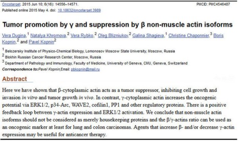
A year ago, in June 2015, Dugina et al published a paper that proposed that altering the ratio of these two actins regulated either suppression or promotion of cancerous cell growth (more work needs to be done). I was very surprised by this idea – even though our work at LPISM had suggested this, I hadn’t thought of putting our observations into the language of “tumor suppression” and “tumor promotion.” Perhaps this was because, in the 1980s, hundreds of so-called “oncogenes” (tumor promoters) and tumor suppressor genes were being cataloged, and our findings were suggesting that a so-called “housekeeping” gene could do the same.

Indeed, Dugina and colleagues even stated this, somewhat simplistically, at the beginning of their Discussion section if their paper:

Until recently non-muscle cytoplasmic β- and γ-actins were considered only to play structural roles in cellular architecture and motility. They (the two isoforms) were viewed as products of housekeeping genes and β-actin was commonly used as internal control in various biochemical experiments.
dugina-citation

It didn’t go unnoticed by me that this paper failed to cite any of our papers, which had produced fundamental knowledge about human cytoplasmic actins. For example, instead of citing our 1980 paper on the amino acid sequences of human cytoplasmic beta- and gamma-actins, the Russian authors cited a paper on the sequences of bovine actins. Furthermore, these authors were apparently unaware of our discovery of actin mutations leading to tumorigenesis and several examples of null alleles of human beta-actin genes associated with tumors.

I communicated by email with the senior author of this paper, Pavel Kopnin at the Blokhin Russian Cancer Research Center in Moscow, not to complain about these omissions, but to tell him that I liked his hypothesis and to explain why. He thanked me and opined that he had had trouble persuading reviewers to publish the paper. I told him that our findings supported his hypothesis and would have made his argument stronger. He apologized for not citing our work and said that he had not reviewed the literature that far back, which amounted to twenty-eight years since our last paper from LPISM was published in 1987 (this made me feel old).

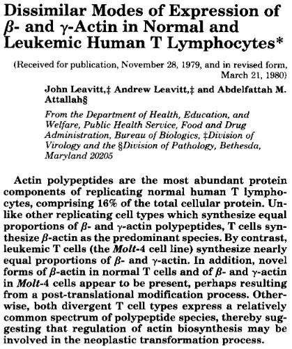
As early as March 1980, I had suggested in writing that altering the ratio of beta- and gamma-actins might contribute to the causation of cancer. This paper was published in the major journal, Journal of Biological Chemistry (see the figure below, last sentence of the abstract). If Dugina et al. were to consider filing a patent on this idea as an invention, our paper would have to, at least, be considered as invalidating prior art along with the rest of our work at LPISM up to 1987.
andy

Both our work at LPISM and Schoenenberger’s work in Basel indicate that the mutation in one of two alleles of the beta-actin gene produces a stable, but defective, form of beta-actin. If Dugina’s hypothesis is correct, it is tempting to suggest that the function of the mutation site in beta-actin controls suppression of tumor formation. I recommended to Pavel Kopnin that his lab pursue this and it is my impression that his lab will continue to work on this hypothesis.

In our model system, we isolated a derivative cell line from the original mutated human fibrosarcoma cell line that exhibited even faster tumor formation (Leavitt et al, 1982). In this second cell line, the mutant beta-actin gene had acquired two additional mutations that made the mutant beta-actin labile with a fast turnover rate in the cell (Lin et al, 1985). As the result of this change, the ratio of stable beta- to gamma-actin changed from approximately 2:1 to approximately 1:1. Furthermore, we found that the two remaining stable forms of beta- and gamma-actin up-regulated in synthesis to maintain a constant normal amount of actin in the cell.

In addition, when we transferred additional mutant human beta-actin genes into immortalized but non-tumorigenic human fibrosarcoma cells, we found that both beta- and gamma-actin from the endogenous normal genes were down-regulated to maintain a constant stable amount of actin in the cell. Thus, we found and reported that beta- and gamma-actin levels in living cells auto-regulated the activities of their own endogenous genes to maintain a constant level of actin in the cell along with a constant ratio of these actins as well (Leavitt et al, 1987a; and Leavitt et al, 1987b). This finding was later confirmed by other laboratories.

These final observations lend support to the idea that maintaining a normal ratio of fully functional cytoplasmic beta- and gamma-actins may be required for the maintenance of the normal, non-neoplastic cellular phenotype. By contrast, mutations and deletions that alter the ratio of functional cytoplasmic beta-actin to gamma actin could lead to tumorigenesis. Hopefully, Pavel Kopnin and others who are aware of our work at LPISM will explore further the role of cytoplasmic actins in maintenance of the normal, non-neoplastic state.

Research Completed at LPISM in 1988 – Reproduced and Extended in 2014

The author in his laboratory at the Linus Pauling Institute of Science and Medicine. Originally published in Science Digest, June 1986.

The author in his laboratory at the Linus Pauling Institute of Science and Medicine. Originally published in Science Digest, June 1986.

[Guest post written by John Leavitt, Ph.D., Nerac, Inc., Tolland, CT.]

In 1987, my colleagues at the Pauling Institute in Palo Alto, colleagues at Stanford and I published a paper that clearly demonstrated that expression of a charge-altered mutant human beta-actin (glycine to aspartic acid substitution at amino acid 245; G245D) caused non-tumorigenic, immortalized human fibroblasts to form aggressive tumors in nude mice (Leavitt et al, 1987a). When these tumor-derived cells were examined, we discovered that they exhibited further elevated expression of the mutant beta-actin and these tumor-derived cells formed tumors even more rapidly – observations that were consistent with the role of this mutation in the tumorigenic phenotype. Furthermore, over-expression of mutant beta-actin was associated with down-regulation of three abundant tropomyosin isoforms in a well-documented transformation-sensitive manner (Leavitt et al, 1986; Leavitt et al, 1987a and Ng et al, 1988). These final papers were the culmination of research conducted at the Linus Pauling Institute of Science and Medicine (LPISM) from December 1981 to March 1988.

Normally when a scientific observation is never repeated it is usually not worth remembering. In this case, twenty-six years after our 1987 publication, a study was published by Schoenenberger et al. at the Biozentrum in Basel, Switzerland, that reproduced our findings in a different cell system, a rat fibroblast model (provided to them by LPISM in 1986). Furthermore, these investigators extended our findings by characterizing new aspects of abnormal behavior of the mutant beta-actin and cells that express this aberrant protein, which help to explain its potential role in cancer such as enhancement of tumor cell motility and invasiveness.

In addition to enhancement of tumor growth and alteration of cell shape, the Swiss investigators presented the following findings to clarify and support the oncogenicity of this mutation:

  1. The mutant actin stimulated formation of ruffles at the cell periphery as shown by staining of cells with an antibody that bound specifically to the mutant epitope of the mutant beta-actin (left image below)
  2. The mutant actin concentrated primarily in these ruffles (palloidin staining reveals the location of filamentous actin in stress fibers; right image below)
  3. The expression of mutant actin inhibited the tropomysin binding to filamentous actin and tropomysin did not accumulate in the ruffles
  4. Mutant actin colocalized with Rac1 (a GTPase mediator of membrane ruffling) and beta1-integrin (adhesion protein) in the ruffles

ruffles

Back-tracking several years, the discovery of this actin mutation was made in a mutagenized cell line isolated by Takeo Kakunaga at the National Cancer Institute (NCI) in 1978. During the month that his paper was published, I walked over to NCI from my lab across the street at the Bureau of Biologics (FDA) to have a chat with Takeo about using his in vitro transformed Syrian Hamster cells as a model system to identify changes in protein expression that correlated with neoplastic transformation. After describing what I wanted to do, he seemed agreeable but then casually mentioned that he had succeed in transforming human fibroblasts into tumor forming cells. I nearly fell off my chair because human cells had never been transformed in vitro before, a major problem for cancer researchers at that time.

I blurted out that we should do the work that I had proposed in his human cell model system, comparing protein expression by the transformed neoplastic cells with their normal precursor cells. My hypothesis was that this comparison would allow identification of proteins that were turned on or turned off in expression by comparing protein profiles of the most abundant 1,000 proteins expressed in these cells and resolved by high-resolution 2-D gel separation (protein profiling). My plan was to look for charge-altering mutations in proteins that might govern neoplastic transformation and tumorigenesis. A fall-back goal was to define the pattern of qualitative and quantitative changes in protein synthesis to try and get a handle on the mysterious mechanism of human cancer development. A summary of the global changes in gene expression of neoplastic human fibroblasts was published from LPISM in 1982 (Leavitt et al, 1982).

Within two weeks, in May of 1978, I was metabolically labeling the total cellular proteins (with the amino acid S-35 methionine) of the normal fibroblasts and three strains of cell lines derived from the normal culture which were immortalized, only one of which formed subcutaneous tumors in nude mice. After four hours of labeling, I prepared extracts of S-35 methionine labeled proteins from each of the four cultures and loaded 25-microliter aliquots of each sample onto the top of clear noodle-like isoelectric focusing gels (7-inch long urea-polyacrylamide gels with the thickness of thin spaghetti) which separated the complex mixture of total cellular proteins by their net charge (isoelectric point). These gels were subjected to isoelectric electrofocusing of the proteins overnight. The next morning I harvested the spaghetti-like gels, and incubated them in a detergent that would bind to the proteins to help separate them by their molecular weights in a second dimension. So, these proteins were first denatured and separated by their net charge and then, in a second dimension, separated by their size on a thin rectangular slab gel.

After about five hours of separation in the second dimension, I was soon to learn that I had separated more than 1,000 denatured protein subunits (polypeptides) by their differing charges and molecular weights. The final step before autoradiography, which revealed the full protein profile, was to fix and stain the gels to get a glimpse of the resolution of these peptide patterns. The staining of these rectangular gels revealed only the most abundant architectural cellular proteins, the largest number of which were cytoplasmic beta- and gamma-actin, at a ratio of about 2:1 in abundance, respectively. The figure below shows what quickly appeared as the gels were de-stained. In the one tumorigenic cell line, instead of seeing a 2:1 ratio of beta- to gamma-actin, a new abundant protein at about one unit charge more negatively charged (more acidic), and half of the normal beta-actin was lost. The pixilation of these three radioactive “spots” immediately suggested to me that one of the two functional genes (alleles) encoding beta-actin had mutated, possibly due to the replacement of a neutral amino acid with a negatively charged amino acid. This prediction was no mystery to me as I had demonstrated this type of electrophoretic shift in another protein a year earlier at Johns Hopkins.

mutant actin further annotated

A number of experiments were done to build the case for the beta-actin mutation, and then I wrote a letter to Klaus Weber at the Max-Planck Institute in Goettingen, Germany, asking for his help in sequencing these actins. His lab was the only one in the world sequencing actins, e.g. the four muscle forms of actins. It only took Klaus two weeks to respond affirmatively, an indication to me that he was eager. I provided him with the actin proteins from this cell line and it took a postdoctoral fellow, Joel Vandekerhkove, and Klaus a little over a year to determine the complete amino acid sequences of the mutant beta-actin and both the wildtype beta- and gamma-actins, to define the mutation that had occurred. We published the result shown above in the top journal Cell in December 1980. Four years later, my colleagues at Stanford and I published my cloning of the mutant and wildtype human beta-actin gene, and the experiments that formally proved the mutation at the level of the gene (Leavitt et al, 1984). Three years after that, we published the experiments that demonstrated the tumorigenic effect of this mutation in immortalized human fibroblasts.

The dramatic nature of this discovery was never fully appreciated, perhaps, because no other actin mutations had been reported and it took Scheonenberger, et al. twenty-six years to complete the work published in September 2013. In another recent related development, Lohr et al. reported reoccurring beta-actin mutations in a panel of tumor cell samples from patients with diffuse large B-cell lymphoma.

One interesting piece of information that came out of our initial sequencing of these actins was the degree of evolutionary conservation of human beta- and gamma-actin. These two actins differ by only four amino acids at the N-terminus, whereas the four muscle-specific human isoforms are more divergent. Comparing the sequence of actin cloned from Saccharomyces cerevisiae (yeast) with these human sequences (sequences stored at the National Center for Biotechnology Information; NCBI) reveals that yeast and human cytoplasmic actins are 92% identical in their sequences (differing by only 31 amino acids out of 375) and most of these amino acid exchanges are conservative replacements both structurally and thermodynamically. This makes these actins the most highly conserved proteins (on a par with histones H3 and H4) among the 20,000 or so known human protein sequences. This fact presents an argument for the fundamental importance of non-muscle cytoplasmic actins in eukaryotic life. It turns out that among actin sequences of all species, no replacement of the Glycine 245 has ever been documented as a result of species divergence or mammalian isoform divergence.

When we introduced the mutant beta-actin gene into a non-tumorigenic immortalized fibroblast strain by gene transfer (Leavitt et al, 1987a), we isolated a transfected clone in which the ratio of exogenous mutant beta-actin to wildtype beta- + gamma-actin was 0.88 – a 76% higher level of expression than the mutant actin in the original mutated cell line in which the mutation arose (0.5 ratio). When we isolated and cultured the cells from a tumor formed by this cell line, the ratio of exogenous mutant beta-actin to wildtype beta- + gamma-actin had increased to 1.95, indicating that about 64% of the total cytoplasmic actin was the mutated beta-actin. Whereas the initial transfectant cell line produced visible tumors at about six weeks, the tumor-derived transfectant cells expressing 64% mutant actin formed visible tumors at about 1.5 weeks. Thus, expression of this mutation was not inhibitory to cell growth.

The other surprising finding was that cell lines expressing the transfected mutant actin gene did not have higher levels of cytoplasmic actins in them because the two endogenous wildtype beta- and gamma-actin genes were coordinately down-regulated (auto-regulated) so that the relative rates of total actin synthesis remained around 30% compared to S-35 methionine incorporation into 600 surrounding non-actin polypeptides in the protein profile (Leavitt et al, 1987b). This auto-regulation phenomenon was reproduced by Minamide et al. (1997) ten years later.

Cytoskeletal rearrangement of actin microfilaments, as well as changes in composition of tropomyosin isoforms and other actin-binding proteins, have long been associated with neoplastic transformation. However, before our study, causal mutations in a cytoplasmic actin had apparently not been considered. It is perhaps consistent then that Ning et al. (2014) have recently described genetically inherited polymorphisms in the actin-bundling protein, plastin (also discovered and cloned at LPISM), that significantly affect the time of tumor recurrence in colorectal cancer after resection and chemotherapy.

During my tenure at the Pauling Institute, I felt that Dr. Pauling understood and appreciated this work and its relevance to the fundamental nature of cancer development. Progress can be slow, but ultimately true understanding of cancer will emerge from this type of research…and I predict that cytoplasmic actins and actin-binding proteins that regulate actin organization and function in the cytoskeleton will be understood to play a central role in the manifestation of the tumorigenic phenotype.

The 1980s at the Linus Pauling Institute – A Wonderful Place to Be

John Leavitt

John Leavitt

[Ed Note: This is part one of a two part series of guest posts written by John Leavitt, Ph.D., Nerac, Inc., Tolland, CT.]

There was an article about Linus Pauling in Time magazine in early 1981 about the fact that at the age of 80 he was still seeking a grant from the National Institutes of Health (NIH) to fund his research on ascorbic acid for treating diseases. This news caught my attention and I looked into the possibility of joining Dr. Pauling’s institute. Toward the end of the summer I was invited to visit the Pauling Institute in Palo Alto, CA to give a seminar on my research at NIH.

In late August Koloman Laki, an aging scientist at NIH, called me up and invited me over to his lab in NIH Building 10, a short walk across the campus from my lab in NIH Building 37. He was interested in talking to me about my recent discovery of mutations in human non-muscle cytoskeletal actin that was published in Cell in late 1980. This protein is the major architectural protein of all eukaryotic cells and we had shown that it was the most highly conserved protein in evolution of the species from yeast to humans. This fact made these mutations even more interesting.

Koloman was a protege of the Hungarian Nobel Prize winner Albert Szent-Györgyi who, I later learned, was much admired by Dr. Pauling because he had discovered both vitamin C and actin. Koloman described how Szent-Györgyi discovered muscle actin. When I mentioned that I was to visit the Linus Pauling Institute in late September, he told me about Emile Zuckerkandl’s and Dr. Pauling’s work on the ‘biological clock,’ which provided evidence in support of Charles Darwin’s theory on divergence of the species.

In the last week of September I flew to Oakland, CA and was picked up at the airport by Emile who was President of the Linus Pauling Institute of Science and Medicine. The next morning I stood up in front of Dr. Pauling and the institute staff to tell them about my discovery of a mutant human beta-actin and my speculation on its involvement in neoplastic transformation. The evidence suggested that I had actually discovered at least two mutations in the same gene, each of which caused a progression to a higher malignant state.

Linus Pauling was in the front row and was all smiles. He asked me if I knew who discovered actin. I was prepared to answer that question thanks to Koloman Laki. In the afternoon I met with Emile who offered me a Senior Scientist position at the Institute, which I accepted. At the time it would be me and Dr. Pauling with separate research interests. Nevertheless, Dr. Pauling could appreciate my discovery as, 32 years earlier, he had described the molecular basis for sickle cell anemia, which predicted that mutations in hemogloblin governed the sickled shape of red blood cells which caused the disease, sickle cell anemia. Likewise, human cancer cells exhibit altered shapes.

So I resigned my secure job-for-life at NIH and moved to Palo Alto to join the struggling Linus Pauling Institute. My technician, Patti Porecca, hired from Bob Gallo’s lab at NIH, would follow me to the Pauling Institute.

Cloning of the Human Beta-Actin Gene

After I arrived at the Pauling Institute, two of my colleagues at NIH and I published a comprehensive study of the changes in protein expression between normal and neoplastic cells in Carcinogenesis using high-resolution computerized microdensitometry to analyze the complex protein patterns (my first paper from the Pauling Institute). This was the first time that such a study had been published, e.g. the comparative profiling of expression of a large number of proteins in neoplastic cells. It was a study of the 1,000 most abundant proteins in normal and neoplastic human cells which revealed potential biomarkers and causative genetic events for human cancer. At the time it was staggering to view these patterns but perfect for my dyslexic brain and mind’s eye. In addition, we published another paper in Cell that described, for the first time, the progression of a neoplastic human cell to a higher malignant cell following a second mutation in the same beta-actin gene. Early in 1982, Steve Burbeck and Jerry Latter at the Institute set up the same computerized microdensitometry platform I had exploited at NIH.

Jerry Latter gave a stirring talk at Argonne Labs in Chicago demonstrating that computerized microdensitometry of protein profiles could be used to determine the identities of unknown proteins based upon determining their amino acid compositions in situ in protein profiles. This paper was published in Clinical Chemistry in 1984. At the same meeting, Steve Burbeck described a truly innovative invention that could measure beta-particles emitted from radioactive protein profiles to produce a direct image of the protein profile pattern. As a group we had entered an exciting period of discovery and innovation at the Linus Pauling Institute.

When I got to Palo Alto in December 1981, I called Professor Larry Kedes at Stanford and we embarked on a collaboration to clone the human beta-actin gene. His impressive postdoctoral fellow, Peter Gunning, taught me some basic recombinant DNA techniques, and I was off to the races. The difficulty was to identify the functional gene in a sea of actin pseudogenes (sometimes referred to as junk DNA). I used an elegant method of homologous recombination developed in Tom Maniatis’ lab at Harvard that had never been used before to clone a novel gene (In fact, cloning of human genes was just getting started at the time). This was smart because Professor Maniatis would be the chairman of the NIH study section that reviewed my first grant proposal submitted from the Pauling Institute. I did not know it at the time but within a month or two I had cloned the functional beta-actin gene a week before Christmas in 1982.

I developed a scheme to identify the correct gene among 300-400 clones of pseudogenes that Patti and I had cloned and the strategy worked. We gave Dr. Sun-Yu Ng the task of sequencing the DNA clone that we were betting on. Rather quickly we determined that we had cloned the functional human beta-actin gene because the DNA sequence that Sun-Yu determined from our candidate clone accurately encoded the amino acid sequence of human beta-actin protein that I had published in Cell in 1980 (with Klaus Weber). Quite coincidentally another lab discovered a rat oncogene that was a fusion of part of an actin gene with a tyrosine kinase gene. I sent this information off to the study section that was reviewing my grant in January 1984 as added evidence that the actin gene was in some way relevant to neoplasia.

My colleagues and I at the Pauling Institute and Stanford published our successful isolation of both the mutant and wildtype human beta-actin genes in Molecular and Cellular Biology in October 1984. As shown below, we had given Armand Hammer’s name to our cancer research program because of his generosity in helping to fund the Linus Pauling Institute.

actin cloning

In January 1984 I was awarded a grant of about $110,000 a year for two years from the American Cancer Society…what a relief. Later in the spring I received word from Professor Maniatis’ NIH study section that our program would also be funded in June by a grant of about $150,000 a year for 3.5 years from the National Cancer Institute for the same work. I was able to hire Dr. Ching Lin from Iowa State University and Dr. Ng (Sun-Yu) from Kedes’ lab. By 1985 Sun-Yu finished the complete DNA sequencing of the human beta-acid gene and Ching sequenced the copy of the beta-actin gene that had two mutations to formally prove the mutations at the level of the gene. Everything that we had learned about the genetic code and amino acid sequences of proteins made our findings predictable. I had learned from my own research how Darwin’s theory of evolution and natural selection worked.

This was the year I finally successfully transferred in recombinant gene inside a cell in culture. I transferred the mutant human actin gene into a rat fibroblast cell line to show that I had cloned the functional gene which could abundantly express its protein the way the natural endogenous beta-actin gene worked (shown in a protein profile below).

mutant actin annotated

At this point I had a brief meeting arranged by Emile with Alex Zafferoni, founder and CEO of Alza Corporation, a block away on Page Mill Road. Zafferoni recommended Bert Roland as a patent attorney. I arranged a meeting with Roland, also a block away, for that afternoon to discuss patenting the human beta-actin gene promoter because of its strong constitutive nature (the engine of the gene that drives its expression). I told Bert that this was a collaboration with Peter Gunning and Larry Kedes at Stanford. Roland was famous for filing Boyer’s and Cohen’s genetic engineering patent which created Genentech and eventually funded Stanford with hundreds of millions of dollars.

We published Sun-Yu’s work on the sequence, structure, and chromosomal location (chromosome 7) of the human beta-actin gene in Molecular and Cellular Biology and we published Ching’s work locating three mutations in this gene in the Proceedings of the National Academy of Sciences, sponsored by Linus Pauling. A patent was filed on the beta-actin promoter and over the years it was licensed to about 15 biotech companies by Stanford University. This patent was prosecuted for the full 17 years (the life of a patent) but never issued. The Institute’s first royalty check was about $10,000 in 1986, but most of the royalties were earned by Stanford’s patent attorneys.

Peter, Larry and I published a paper in PNAS on the use of the human beta-actin gene promoter for expression of other genes. This vector was distributed to anyone who asked for it – and many did – and to those companies that licensed the invention. At last count this paper had more than 1,000 reference citations.

Our paper popularized the actin promoter as a strong constitutive promoter of foreign gene expression. Soon the rice actin promoter would be used to make Round-up Ready crops by DeKalb Genetics and Monsanto, and giant tilapia fish would be engineered with growth hormone under the control of the fish beta-actin promoter. There were even fluorescent mice running around in Japan created with firefly luciferase expressed by the beta-actin promoter (which I called “the cat’s meow”). Since cytoplasmic actins are the most abundant proteins in most cells you could use the promoter to abundantly express foreign genes in most cells of any animal.

In 1987 we also published the culmination of my research on the mutant beta-actin gene in Molecular and Cellular Biology. When I introduced this gene into non-tumor forming immortalized human fibroblasts they became tumorigenic. The results showed that the more abundant the expression of the mutant beta-actin, the more tumorigenic the non-tumorigenic cells became and the cells that came out of the tumors were enhanced further in the level of mutant beta-actin expression. This was a sensational finding that was the goal of research which began with the discovery of the mutant beta-actin in 1978 at NIH.